. 2017 Nov 2;551(7678):110-114.
doi: 10.1038/nature24293. Epub 2017 Oct 25.

IL-1R8 is a checkpoint in NK cells regulating anti-tumour and anti-viral activity

Affiliations

IL-1R8 is a checkpoint in NK cells regulating anti-tumour and anti-viral activity

Martina Molgora et al. Nature. .

Abstract

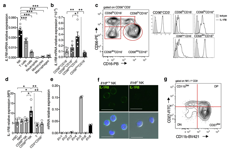
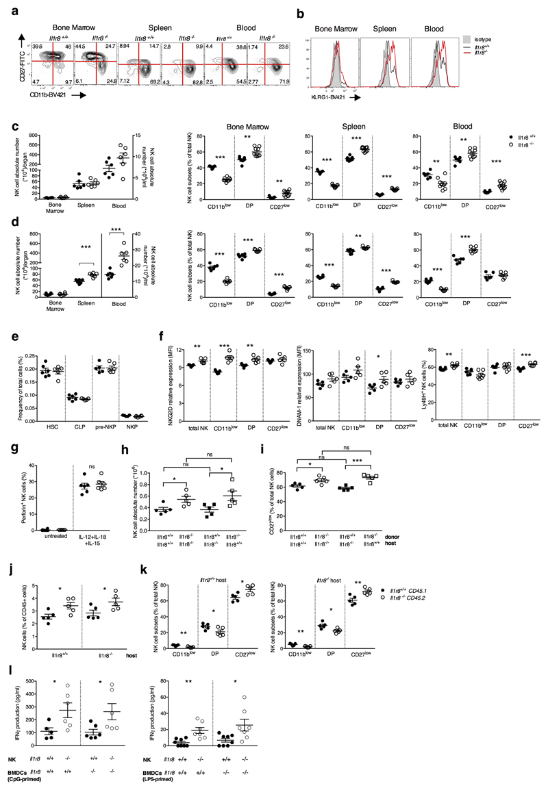
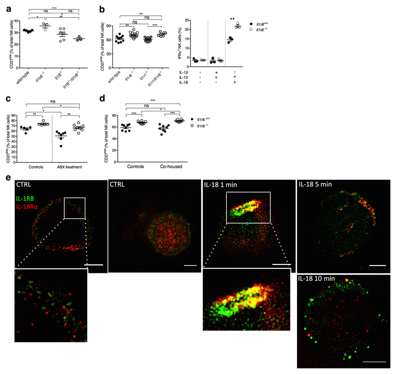
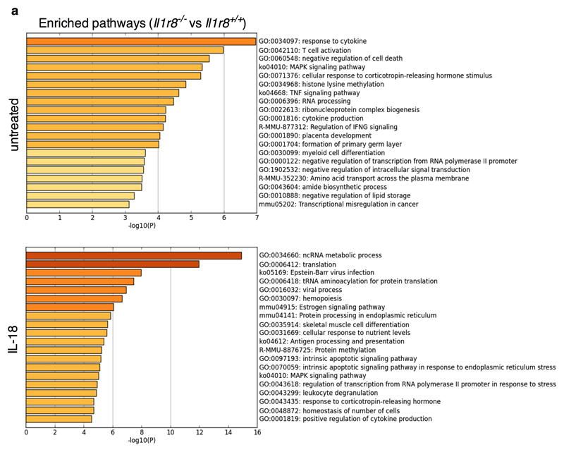
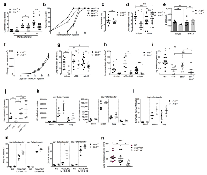
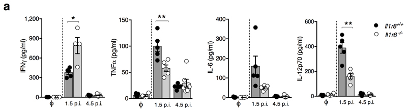
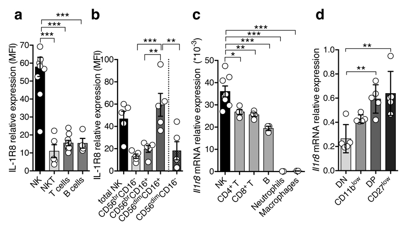
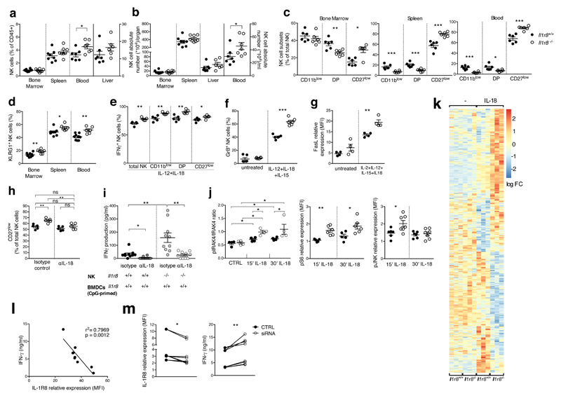
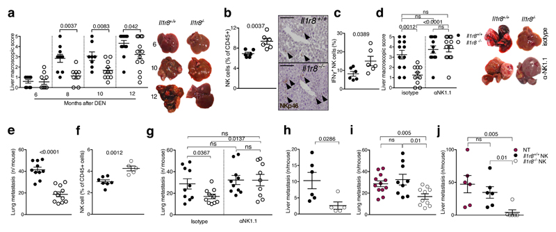
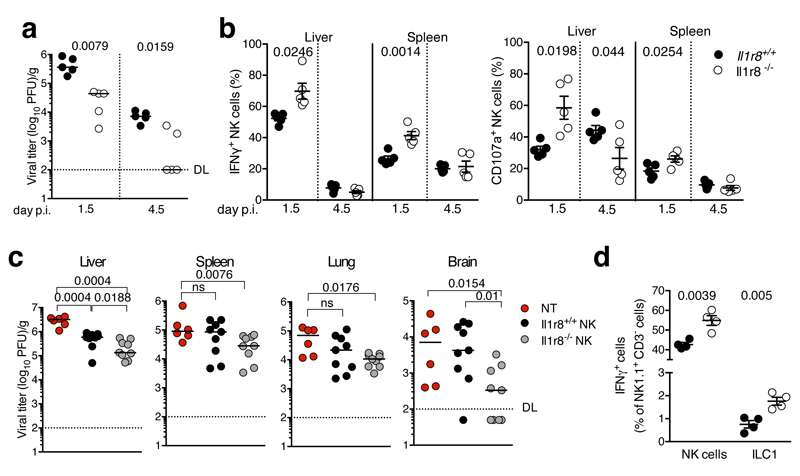
Interleukin-1 receptor 8 (IL-1R8, also known as single immunoglobulin IL-1R-related receptor, SIGIRR, or TIR8) is a member of the IL-1 receptor (ILR) family with distinct structural and functional characteristics, acting as a negative regulator of ILR and Toll-like receptor (TLR) downstream signalling pathways and inflammation. Natural killer (NK) cells are innate lymphoid cells which mediate resistance against pathogens and contribute to the activation and orientation of adaptive immune responses. NK cells mediate resistance against haematopoietic neoplasms but are generally considered to play a minor role in solid tumour carcinogenesis. Here we report that IL-1R8 serves as a checkpoint for NK cell maturation and effector function. Its genetic blockade unleashes NK-cell-mediated resistance to hepatic carcinogenesis, haematogenous liver and lung metastasis, and cytomegalovirus infection.


Conflict of interest statement

Authors declare no Competing financial interests.

Figures

Extended Data Figure 1
Extended Data Figure 1. Expression of IL-1R8 in human and murine NK cells
(a, b) IL-1R8 mRNA (a) expression in human primary NK cells, compared with T and B cells, neutrophils, monocytes and in vitro-derived macrophages (a) and in human primary NK cell maturation stages (CD56brCD16-, CD56brCD16+, CD56dimCD16+), and in the CD56dimCD16- subset (b). (c) Representative FACS plot of human NK cell subsets and histograms of IL-1R8 expression in NK cell subsets. (d) IL-1R8 protein expression in human bone marrow precursors and mature cells. (e) IL-1 receptor family members (Il1r1, Il1r2, Il1r3, Il1r4, Il1r5, Il1r6, Il1r8) mRNA expression in murine primary NK cells isolated from the spleen. (f) IL-1R8 protein expression in murine NK cells by confocal microscopy. Magnification bar: 10μm. (g) Representative FACS plot of murine NK cell subsets. (a, b, d) *p < 0.05, **p < 0.01, ***p < 0.001 One-way ANOVA. Mean ± SEM. a, n=6 (NK and B cells) or n=4 donors; b, n=5 donors; d, n=4 donors; e, n=2 mice; f, representative images out of four collected per group. a, b, d, e, f, one experiment performed.
Extended Data Figure 2
Extended Data Figure 2. Phenotypic analysis of Il1r8-/- NK cells.
(a, b) Representative FACS plot of murine NK cell subsets in Il1r8+/+ and Il1r8-/- mice (a) and histograms of KLRG1 expression in NK cells. (c, d) NK absolute number and NK cell subsets (DN, CD11blow, DP and CD27low) in bone marrow, spleen and blood of Il1r8+/+ and Il1r8-/- newborn mice at 2 (c) and 3 (d) weeks of age. (e) Frequency of bone marrow precursors in Il1r8+/+ and Il1r8-/- mice. (f) NKG2D, DNAM-1 and LY49H expression in peripheral NK cells and NK cell subsets of Il1r8+/+ and Il1r8-/- mice. (g) Frequency of splenic Perforin+ NK cell subsets upon stimulation in Il1r8+/+ and Il1r8-/- mice. (h and i) Peripheral NK cell absolute number (h) and CD27low NK cell frequency (i) in bone marrow chimeric mice upon reconstitution (9 weeks). (j and k) Peripheral NK cell (j) and NK cell subset (k) frequency in competitive chimeric mice transplanted with 50% of Il1r8+/+ CD45.1 cells and 50% of Il1r8-/- CD45.2 cells upon reconstitution (9 weeks). Upon reconstitution a defective engraftment (12% instead of 50% engraftment) of Il1r8-/- stem cells was observed in competitive conditions. (l) IFNγ production by Il1r8+/+ and Il1r8-/- NK cells upon co-culture with LPS- or CpG-primed Il1r8+/+ and Il1r8-/-DCs. (c-l) *p < 0.05, **p < 0.01, ***p < 0.001 between selected relevant comparisons, two-tailed unpaired Student’s t test. Centre values and error bars represent mean ± SEM. At least 5 animals per group were used. c, d: 3 pooled experiments. e-l: one experiment was performed.
Extended Data Figure 3
Extended Data Figure 3. Mechanism of IL-1R8-dependent regulation of NK cells
(a) Splenic CD27low NK cell frequency in wild type, Il1r8-/-, Il18-/-, and Il18-/-/Il1r8-/- mice. (b) Peripheral CD27low NK cell frequency in wild-type, Il1r8-/-, Il1r1-/- and Il1r8-/-Il1r1-/- mice (left) and IFNγ production by splenic NK cells after IL-12 and IL-1β or IL-18 stimulation (right). (c, d) Splenic CD27low NK cell frequency in Il1r8+/+ and Il1r8-/- mice upon commensal flora depletion (c) and breeding in co-housing conditions (d). (e) STED microscopy of human NK cells stimulated with IL-18. Magnification bar: 2μm. (a-d) *p < 0.05, **p < 0.01, ***p < 0.001 between selected relevant comparisons, two-tailed unpaired Student’s t test; Centre values and error bars represent mean ± SEM. a, n= 3, 5, or 6 mice; at least 5 animals per group were used (b-d). a-d: one experiment was performed. e: representative images out of three collected from two donors.
Extended Data Figure 4
Extended Data Figure 4. RNA-seq analysis of Il1r8+/+ and Il1r8-/- NK cells.
(a) Metascape analysis of enriched gene pathways of resting and IL-18-activated Il1r8+/+ and Il1r8-/- NK cells. See also Supplementary Table 1 and data deposited in NCBI Gene Expression Omnibus accessible through GEO Series accession number GSEXXXXX.
Extended Data Figure 5
Extended Data Figure 5. NK cell-mediated resistance to HCC and metastasis in IL-1R8-deficient mice.
(a) Macroscopic score of liver lesions in female Il1r8+/+ and Il1r8-/- mice 6, 10 and 12 months after DEN injection. (b) HCC incidence in Il1r8+/+ and Il1r8-/- female and male mice. (c) Frequency of IFNγ+ NK cells in spleen of Il1r8+/+ and Il1r8-/- tumor bearing mice. (d) Macroscopic score of liver lesions in female Il1r8+/+ and Il1r8-/- mice upon NK cell depletion. (e) 2-DG quantification in lungs of Il1r8+/+ and Il1r8-/- tumor bearing mice upon NK cell depletion. (f) Primary tumor growth in Il1r8+/+ and Il1r8-/- mice (25 days after MN/MCA1 cell line injection). (g) Number of lung metastasis in Il1r8+/+ and Il1r8-/- MN/MCA1 sarcoma bearing mice upon IFNγ or IL-18 neutralization. (h) Volume of lung metastases in Il1r8+/+ and Il1r8-/- MN/MCA1-bearing mice upon depletion of IL-17A or CD4+/CD8+ cells. (i) Number of lung metastases in Il1r8+/+ and Il1r8-/-, Il1r1-/-, Il1r1-/-/Il1r8-/- MN/MCA1-bearing mice. (j) Number of liver metastasis in Il1r8+/+, Il1r8-/-, Il18-/-, Il18-/-Il1r8-/- MC38 colon carcinoma bearing mice. (k) Il1r8+/+ and Il1r8-/- NK cell absolute number three or 7 days after adoptive transfer. (l) In vivo Il1r8+/+ and Il1r8-/- NK cell proliferation three days after adoptive transfer. (m) Ex vivo IFNγ production and degranulation upon 4-hour stimulation with PMA-Ionomycin, IL-12 and IL-18 in adoptively transferred Il1r8+/+ and Il1r8-/- NK cells. (n) Volume of lung metastasis of Il1r8+/+ MN/MCA1 sarcoma bearing mice after adoptive transfer of Il1r8+/+ and Il1r8-/- NK cells. (a, c-e, g-j, m-n) *p < 0.05, **p < 0.01, ***p < 0.001 between selected relevant comparisons, two-tailed unpaired Student’s t test or Mann-Whitney test. #p<0.05, ##p<0.01, Kruskal-Wallis and Dunn's multiple comparison test. Centre values and error bars represent mean ± SEM. a, n=9, 10, 11, 18, 21 mice; b, n=8-21 mice; c, n=6 mice; d, n= 10, 12, 13 mice; e, n=4 (Il1r8-/- isotype) or n=5; f, n=10; g, n=6, 7, 9, 10 mice; h, n=5, 6, 12 mice; i, n=6, 8, 10 mice; j, n=4, 5, 7 mice; k, l, m, n=3 mice; n, n=9, 10, 12 mice. Representative experiment out of 3 (a, b), 2 (d), 6 (f), or one (c, e, g-n) experiment performed.
Extended Data Figure 6
Extended Data Figure 6. NK cell-mediated antiviral resistance in IL-1R8-deficient mice
(a) Cytokine serum levels in Il1r8+/+ and Il1r8-/- infected mice (1.5 and 4.5 days post infection). *p < 0.05, **p < 0.01, ***p < 0.001, unpaired Student’s t test. Centre values and error bars represent mean ± SEM. n=5 mice. One experiment was performed.
Figure 1
Figure 1. Expression of IL-1R8 in human and murine NK cells
(a, b) IL-1R8 protein expression in human primary NK cells and other leukocytes (a) and NK cell maturation stages (b). (c, d) Il-1r8 mRNA expression in murine primary NK cells and other leukocytes (c) and in sorted splenic NK cell subsets (c). *p < 0.05, **p < 0.01, ***p < 0.001 One-way ANOVA. Mean ± SEM.
Figure 2
Figure 2. NK cell differentiation and function in IL-1R8-deficient mice
(a, b) NK cell frequency and absolute number among leukocytes in Il1r8+/+and Il1r8-/- mice. (c, d) NK cell subsets (c) and KLRG1+ NK cells (d). (e-g) IFNγ (e), Granzyme B (f) and FasL (g) expression in stimulated NK cells. (h) Splenic CD27low NK cell frequency upon IL-18 in vivo depletion. (i) IFNγ production by Il1r8+/+ and Il1r8-/- NK cells upon co-culture with CpG-primed Il1r8+/+ DCs and IL-18 blockade. (j) IRAK4, S6 and JNK phosphorylation in NK cells upon stimulation with IL-18. (k) RNA-seq analysis of resting and IL-18-activated NK cells. Differentially expressed (p<0.05) genes are shown. FC: fold change. (l) Correlation between IL-1R8 expression and IFNγ production in human peripheral blood NK cells. (m) IL-1R8 expression and IFNγ production in human NK cells 7 days after transfection with control siRNA or IL-1R8-specific siRNA in duplicate. (a-l) *p < 0.05, **p < 0.01, ***p < 0.001 between selected relevant comparisons, two-tailed unpaired Student’s t test or Mann-Whitney test; (k) r: Pearson correlation coefficient; Mean ± SEM.
Figure 3
Figure 3. NK cell-mediated protection against liver carcinogenesis and metastasis in IL-1R8-deficient mice
(a) Macroscopic score of liver lesions in male Il1r8+/+ and Il1r8-/- mice 6, 8, 10 and 12 months after DEN injection. (b) Frequency and representative histological quantification of NK cell infiltrate in liver of tumor bearing mice. (20X, bar = 100µm). (c) Frequency of IFNγ+ NK cells in liver of tumor bearing mice. (d) Macroscopic score of liver lesions in male mice upon NK cell depletion. (e) Number of spontaneous lung metastasis. (f) NK cell frequency in the lungs of MN/MCA1 tumor bearing mice. (g) Number of lung metastasis in MN/MCA1 tumor bearing mice upon NK cell depletion. (h) Number of liver metastasis in MC38 colon carcinoma bearing mice. (i,j) Number of lung (i) and liver (j) metastasis of Il1r8+/+ mice after adoptive transfer of Il1r8+/+ and Il1r8-/- NK cells. (a, d) Representative images of female livers are shown. (a-j) *p < 0.05, **p < 0.01, ***p < 0.001 between selected relevant comparisons, two-tailed unpaired Student’s t test. Mean ± SEM.
Figure 4
Figure 4. NK cell-mediated antiviral resistance in IL-1R8-deficient mice
(a) Viral titer in livers of Il1r8+/+ and Il1r8-/- infected mice. (b) Frequency of IFNγ+ and CD107a+ NK cells of infected mice. (c) Viral titers in newborn wild type mice upon adoptive transfer of Il1r8+/+ and Il1r8-/- NK cells (7 days post infection). (d) Frequency of IFNγ + cells in the liver of MCMV infected mice. (a-d) *p < 0.05, **p < 0.01, ***p < 0.001, two-tailed Mann-Whitney test (a, c) or unpaired Student’s t test (b, d). Median (a, c). Mean ± SEM (b, d). DL: detection limit. Day p.i.: day post infection.

Comment in

  • A new checkpoint for Natural Killer cell activation.
    Souza-Fonseca-Guimaraes F, Huntington ND. Souza-Fonseca-Guimaraes F, et al. Immunol Cell Biol. 2018 Jan;96(1):5-7. doi: 10.1111/imcb.1027. Epub 2017 Dec 15. Immunol Cell Biol. 2018. PMID: 29356093 No abstract available.

References

    1. Garlanda C, Dinarello CA, Mantovani A. The interleukin-1 family: back to the future. Immunity. 2013;39:1003–1018. - PMC - PubMed
    1. Di Santo JP. Natural killer cell developmental pathways: a question of balance. Annu Rev Immunol. 2006;24:257–286. - PubMed
    1. Vivier E, et al. Innate or adaptive immunity? The example of natural killer cells. Science. 2011;331:44–49. - PMC - PubMed
    1. Bellora F, et al. Human NK cells and NK receptors. Immunol Lett. 2014;161:168–173. - PubMed
    1. Guillerey C, Huntington ND, Smyth MJ. Targeting natural killer cells in cancer immunotherapy. Nat Immunol. 2016;17:1025–1036. - PubMed

Publication types

MeSH terms