Review
. 2017 Oct 26:3:17077.
doi: 10.1038/nrdp.2017.77.

Merkel cell carcinoma

Affiliations
Review

Merkel cell carcinoma

Jürgen C Becker et al. Nat Rev Dis Primers. .

Abstract

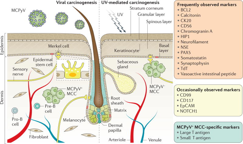
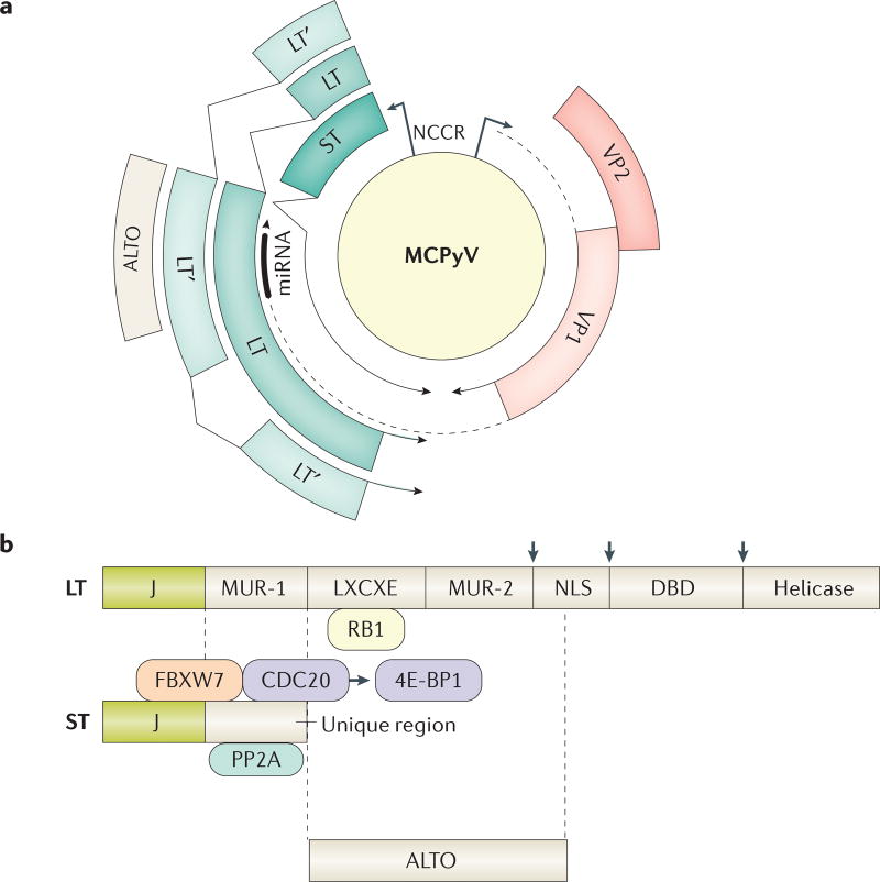
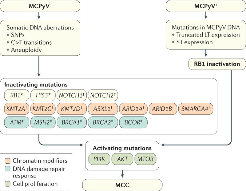
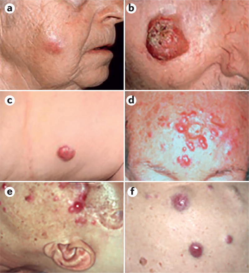
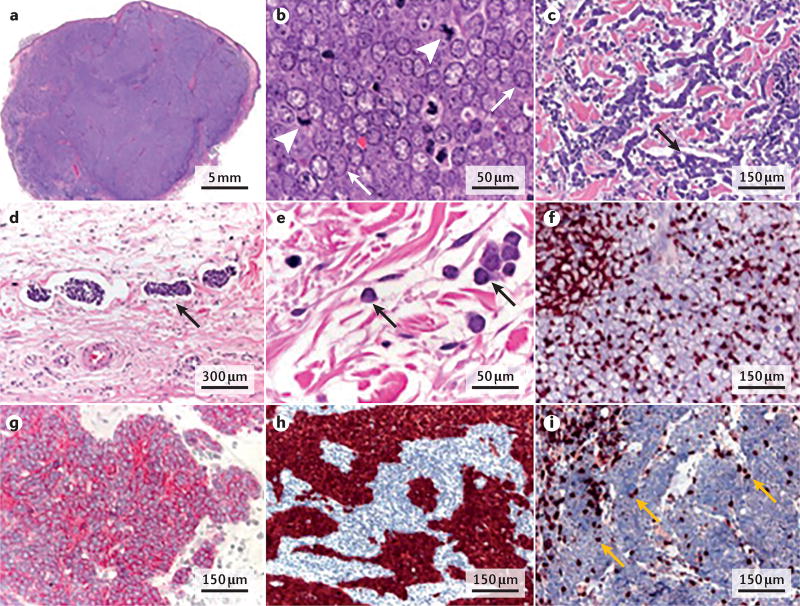
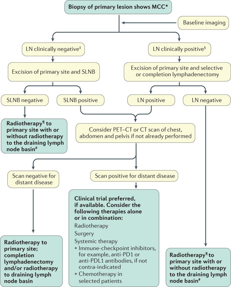
Merkel cell carcinoma (MCC) is a rare but highly aggressive skin cancer with neuroendocrine features. MCC pathogenesis is associated with either the presence of Merkel cell polyomavirus or chronic exposure to ultraviolet light (UV), which can cause a characteristic pattern of multiple DNA mutations. Notably, in the Northern hemisphere, the majority of MCC cases are of viral aetiology; by contrast, in areas with high UV exposure, UV-mediated carcinogenesis is predominant. The two aetiologies share similar clinical, histopathological and prognostic characteristics. MCC presents with a solitary cutaneous or subcutaneous nodule, most frequently in sun-exposed areas. In fact, UV exposure is probably involved in both viral-mediated and non-viral-mediated carcinogenesis, by contributing to immunosuppression or DNA damage, respectively. Confirmation of diagnosis relies on analyses of histological features and immunological marker expression profiles of the lesion. At primary diagnosis, loco-regional metastases are already present in ∼30% of patients. Excision of the tumour is the first-line therapy; if not feasible, radiotherapy can often effectively control the disease. Chemotherapy was the only alternative in advanced-stage or refractory MCC until several clinical trials demonstrated the efficacy of immune-checkpoint inhibitors.


Conflict of interest statement

Competing interests statement

A.S., L.C. and M.V. declare no competing interests.

Figures

Figure 1
Figure 1. Hypothetical cells of origin, causal events and tissue markers for MCC
The cell of origin of Merkel cell carcinoma (MCC) has not been identified. Possible candidates include epidermal stem cells, keratinocytes (the predominant cells in all the epidermal cell layers), dermal fibroblasts, pro-B cells or pre-B cells. Fibroblasts, pro-B cells and pre-B cells are localized in the dermal compartment, which is not exposed to relevant amounts of ultraviolet light (UV), and, therefore, are probably not cells of origin in UV-mediated carcinogenesis. Merkel cells are postmitotic cells and, therefore, are probably not the cell of origin of MCC. Merkel cells are found in the basal layer of the epidermis and are probably derived from epidermal or hair follicle stem cells. Merkel cells function as mechanoreceptors to detect gentle touch and are associated with sensory nerves. Merkel cell polyomavirus (MCPyV) is a common component of the commensal skin microbiota. However, it is not known what cell type MCPyV preferentially infects. In countries with low UV exposure, the majority of MCCs are positive for MCPyV (MCPyV+ MCC), whereas in countries with high UV exposure, MCPyV is less frequently associated with MCC; these MCPyV MCCs are characterized by DNA mutations bearing a UV signature. The two MCC types have similar phenotypes. Tissue markers that can be frequently or occasionally observed in both MCPyV+ MCC and MCPyV MCC, as well as MCPyV+ MCC-specific markers, are listed. BCL2, apoptosis regulator BCL2; CK20, cytokeratin 20; CD56, neural cell adhesion molecule 1; CD99, CD99 antigen; CD117, mast/stem cell growth factor receptor Kit; EpCAM, epithelial cell adhesion molecule; HIP1, huntingtin-interacting protein 1; NSE, neuron-specific enolase, also known as γ-enolase; NOTCH1, neurogenic locus notch homologue protein 1; PAX5, paired box protein Pax-5; TdT, DNA nucleotidylexotransferase.
Figure 2
Figure 2. Circular map of MCPyV and linear maps of the MCPyV early genes
a | Merkel cell polyomavirus (MCPyV) has a 5,387 bp circular double-stranded DNA genome with two transcriptional units, the early and late regions. The early region yields four spliced mRNAs encoding four proteins: two alternatively spliced isoforms of the large T antigen (LT and LT’, which is also known as 57 kT), the small T antigen (ST) and ALTO (alternate frame of the LT open reading frame). The late region encodes two viral coat proteins, VP1 and VP2, and a microRNA that targets the T antigen transcripts,,. b | LT contains an N-terminal J domain, MCPyV-unique region (MUR)-1 and MUR-2, LXCXE motif (where the retinoblastoma-associated protein (RB1) binds), nuclear localization signal (NLS), DNA or origin binding domain (DBD) and helicase domain. The cell growth-inhibitory domain (not shown) overlaps with the helicase domain. On the basis of its similarity to other polyomaviruses, MCPyV LT is thought to form two hexamers that bind in head-to-head fashion to the origin of replication and serves to melt, twist and unwind the viral DNA and recruit the cellular DNA polymerases of the host cell to enable viral replication. Which cells normally support MCPyV replication in humans is unknown, as MCPyV LT expression has not yet been detected by immunohistochemistry in any normal human tissue. However, cultures of primary human dermal fibroblasts could support MCPyV replication. In Merkel cell carcinoma (MCC), mutations in MCPyV DNA result in truncated LTs (indicated by arrows) that retain the LXCXE motif and sometimes the NLS and can bind and inhibit RB1. ST contains an N-terminal J domain and a unique domain not shared with LT. ST can bind to regulatory and catalytic subunits of protein phosphatase 2A (PP2A). The LT stabilizing domain (not shown) within the unique domain is distinct from the sequence that binds the phosphatase and participates in binding to F-box/WD repeat-containing protein 7 (FBXW7) and cell division cycle protein 20 homologue (CDC20). MCPyV ST binding to CDC20 could contribute to increased phosphorylation of eukaryotic translation initiation factor 4E-binding protein 1 (4E-BP1). NCCR, non-coding control region.
Figure 3
Figure 3. Genetic aberrations in MCC
Merkel cell carcinoma (MCC) develops from substantial changes in the genome that originate from ultraviolet light damage (including point mutations, amplifications, deletions and translocations) or integration of the Merkel cell polyomavirus (MCPyV) genome and expression of large T antigen (LT) and small T antigen (ST) that lead to perturbations in a variety of signalling pathways. The retinoblastoma-associated protein (RB1) pathway, which normally has tumour-suppressive roles, is altered by mutations in RB1 in MCPyV MCC and by LT in MCPyV+ MCC, which perturbs the ability of RB1 to inhibit transcription factors of the E2F family. Cellular tumour antigen p53 (encoded by TP53) contributes to regulating the cell cycle by activating genes that negatively regulate cell division; NOTCH1 and NOTCH2 encode receptors involved in cell differentiation and proliferation. SNPs, single-nucleotide polymorphisms. *Mutation observed in most cases; mutation observed frequently.
Figure 4
Figure 4. Clinical presentations of MCC
a | Cutaneous–subcutaneous nodule on sun-exposed skin of an elderly woman. b | Large, partly ulcerated tumour on sun-exposed skin of an elderly man. c | Small cutaneous tumour on the thigh of an immunosuppressed woman. d | Satellite metastases on the forehead of an elderly woman. e | In-transit metastases on the face of an immunocompromised woman. f | Multiple cutaneous distant metastases on the back of a woman.
Figure 5
Figure 5. Histopathological and immunohistochemical features of MCC
Small-blue-round-cell tumours such as Merkel cell carcinoma (MCC) owe their name to the colour of the cancerous cells after haematoxylin and eosin staining. a | Large dermal and subcutaneous nodule. b | Higher-magnification view of the tissue in part a shows monomorphic mid-sized cells with vesicular nuclei, scanty cytoplasm (arrows) and several mitoses (arrowheads). c | The trabecular pattern characterized by anastomosing (connecting) cords of tumour cells (arrow) was the feature that gave MCC its first name of trabecular carcinoma of the skin. This feature is relatively uncommon and is usually found at the periphery of the tumour. d | Intra-lymphatic complexes of tumour cells (arrow). e | Isolated tumour cells near the margin of the surgical excision (arrows). f | Strong positivity for cytokeratin 20 (CK20) staining (brown), with a dot-like perinuclear accentuation, although a more-diffuse cytoplasmic pattern can also be observed. g | Positivity for synaptophysin (dark pink). h | Strong positivity for the Merkel cell polyomavirus (MCPyV) large T antigen (brownish red). i | Staining for CD8, which reveals some intratumoural (arrows) and several peritumoural CD8+ T lymphocytes.
Figure 6
Figure 6. Simplified evaluation and treatment of primary MCC
Algorithm for diagnostic and therapeutic decisions for managing patients with Merkel cell carcinoma (MCC). The flowchart begins with the assessment of the extent of disease spread to distant sites (baseline imaging) and regional nodal disease (typically including pathological assessment of clinically negative nodes). After staging is complete, the appropriate therapy can be identified. See the National Comprehensive Cancer Network guidelines and http://www.merkelcell.org/ for further information, including surveillance guidance. LN, lymph node; PD1, programmed cell death protein 1; PDL1, PD1 ligand 1; SLNB, sentinel lymph node biopsy. *Consider baseline Merkel cell polyomavirus serology for prognostic significance and to track disease. No pathologically enlarged nodes on physical examination and by imaging study. §Pathologically enlarged nodes on physical examination or by imaging study. Radiotherapy is indicated in most patients, with the exception of low-risk disease (for example, primary tumour ≤1 cm on the extremities or trunk, no lymphovascular invasion or negative surgical margin) in patients who are not immunosuppressed. #Consider radiotherapy to the nodal basin in high-risk patients.

Similar articles

Cited by

References

    1. Toker C. Trabecular carcinoma of the skin. Arch. Dermatol. 1972;105:107–110. - PubMed
    1. Maricich SM, et al. Merkel cells are essential for light-touch responses. Science. 2009;324:1580–1582. This study demonstrates that Merkel cells are essential for gentle touch reception. - PMC - PubMed
    1. Lemos BD, et al. Pathologic nodal evaluation improves prognostic accuracy in Merkel cell carcinoma: analysis of 5823 cases as the basis of the first consensus staging system. J. Am. Acad. Dermatol. 2010;63:751–761. - PMC - PubMed
    1. Schrama D, et al. Merkel cell polyomavirus status is not associated with clinical course of Merkel cell carcinoma. J. Invest. Dermatol. 2011;131:1631–1638. - PubMed
    1. Becker JC, Schrama D, zur Hausen A. In: Rook’s Textbook of Dermatology. 9. Ch. 145. Griffiths C, Barker J, Bleiker T, Chalmers R, Creamer D, editors. Vol. 12. John Wiley & Sons, Inc.; 2016.

MeSH terms