. 2017 Oct 24;114(43):11404-11409.
doi: 10.1073/pnas.1706617114. Epub 2017 Oct 9.

Human SHMT inhibitors reveal defective glycine import as a targetable metabolic vulnerability of diffuse large B-cell lymphoma

Affiliations

Human SHMT inhibitors reveal defective glycine import as a targetable metabolic vulnerability of diffuse large B-cell lymphoma

Gregory S Ducker et al. Proc Natl Acad Sci U S A. .

Abstract

The enzyme serine hydroxymethyltransferse (SHMT) converts serine into glycine and a tetrahydrofolate-bound one-carbon unit. Folate one-carbon units support purine and thymidine synthesis, and thus cell growth. Mammals have both cytosolic SHMT1 and mitochondrial SHMT2, with the mitochondrial isozyme strongly up-regulated in cancer. Here we show genetically that dual SHMT1/2 knockout blocks HCT-116 colon cancer tumor xenograft formation. Building from a pyrazolopyran scaffold that inhibits plant SHMT, we identify small-molecule dual inhibitors of human SHMT1/2 (biochemical IC50 ∼ 10 nM). Metabolomics and isotope tracer studies demonstrate effective cellular target engagement. A cancer cell-line screen revealed that B-cell lines are particularly sensitive to SHMT inhibition. The one-carbon donor formate generally rescues cells from SHMT inhibition, but paradoxically increases the inhibitor's cytotoxicity in diffuse large B-cell lymphoma (DLBCL). We show that this effect is rooted in defective glycine uptake in DLBCL cell lines, rendering them uniquely dependent upon SHMT enzymatic activity to meet glycine demand. Thus, defective glycine import is a targetable metabolic deficiency of DLBCL.

Keywords: DLBCL; SHMT; cancer metabolism; folate; glycine.


Conflict of interest statement

Conflict of interest statement: N.M., V.S., A.F., and M.G.M. are employees of Raze Therapeutics. J.D.R. is a founder and member of the scientific advisory board of Raze Therapeutics. G.S.D., J.M.G., H.K., and J.D.R. are inventors on a Princeton University patent covering serine hydroxymethyltransferse inhibitors and their use in cancer.

Figures

Fig. 1.
Fig. 1.
SHMT is required for tumor formation in vivo. (A) Serine synthesis and catabolism occur in an intercompartmental cycle mediated by cytosolic and mitochondrial SHMT activity. Key enzymes mediating these transformations are highlighted in capital letters. Arrows indicate the directionality of flux in HCT-116 cells, but most reactions are readily reversible. (B) Growth of subcutaneous tumors from HCT-116 WT and ΔSHMT2 cells implanted in opposite flanks of nude mice (mean ± SEM, n = 10, *P < 0.05, paired t test). (C) Tumor growth of subcutaneous tumors from HCT-116 ΔSHMT2 and ΔSHMT1/2 cells implanted in opposite flanks of nude mice (mean ± SEM, n = 10).
Fig. S1.
Fig. S1.
SHMT1/2 deletion cell lines cannot establish xenografts in nude mice. (A) Weights of mice from experiments shown in Fig. 1 B and C (mean ± SD, n = 10). (B) Intratumor abundance of AICAR and serine from xenografted tumors (mean ± SD, n = 9, ***P < 0.001, paired t test). (C) Western blot showing loss of SHMT1 and SHMT2 expression in SHMT1/2 double-deletion HCT-116 cell lines. (D) Growth of HCT-116 WT and SHMT1/2 double-deletion cells in standard DMEM with and without supplemental 1 mM sodium formate (mean ± SD, n ≥ 4). (E) Representative mice 35 d after injection with HCT-116 SHMT2 deletion cells (right flank) or HCT-116 SHMT1/2 double-deletion cells (left flank).
Fig. 2.
Fig. 2.
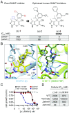
A folate-competitive cell-permeable inhibitor of human SHMT1/2. (A) Structure of pyrazolopyran inhibitor of plant SHMT (compound 1) and two optimized inhibitors for human SHMT1/2 (compounds 2 and 3; compound 3 = SHIN1). IC50s shown are for human SHMT1 and -2 in an in vitro assay. (B, Left) Compound 2 in complex with human SHMT2 as solved in a 2.5-Å resolution X-ray crystal structure. The electron density of the compound is shown as the 2Fo–Fc map contoured at 0.5 σ and generated with compound 2 omitted. (Right) An overlay of the SHMT2/compound 2 structure with the structure of 5-formyl–THF-triglutamate in complex with rabbit SHMT1. (C) Growth of HCT-116 WT ±1 mM formate and ΔSHMT1 and ΔSHMT2 cell lines in the presence of increasing concentrations of SHIN1 (n ≥ 3). (D) Cellular IC50 values for growth inhibition by compound 2 and SHIN1.
Fig. S2.
Fig. S2.
A folate competitive SHMT inhibitor. (A) Enzymatic inhibition of human SHMT1 and SHMT2 with enantiomerically resolved fractions of compound 2. (B) (−)-SHIN1 does not inhibit growth of HCT-116 cells at concentrations up to 30 µM (mean ± SD, n = 3). (C) Formate rescue of cell growth in SHIN1 treated cells requires glycine. Normalized growth of HCT-116 cells treated with 10 µM (+)-SHIN1 in DMEM with and without standard glycine and supplemented with formate (mean ± SD, n = 3). (D) Growth of human pancreatic cell line 8988T with indicated concentrations of SHIN1 (mean ± SD, n = 6).
Fig. 3.
Fig. 3.
(+)-SHIN1 inhibits SHMT1/2 in HCT-116 cells. (A) M+2 13C-labeling fraction of intracellular ADP and glutathione after 24 h 13C-serine coincubation with DMSO, 5 µM (+)-SHIN1, or 5 μM (–)-SHIN1. ΔSHMT1/2 cells were cultured without formate for the duration of labeling (mean ± SD, n = 3). (B) Normalized (to DMSO HCT-116 WT cells) levels of purine biosynthetic pathway intermediates after 24-h incubation ±SHIN1 (mean ± SD, n = 3). (C) Total metabolite abundances in HCT-116 cells treated with DMSO vs. (+)-SHIN1 (10 µM) for 48 h. Metabolites whose abundances differ by more than fourfold between conditions are highlighted in red (mean, n = 3). (D) Metabolite abundance in HCT-116 cells treated with DMSO or SHIN1 in the presence of 1 mM sodium formate. The same metabolites whose abundances were different in C are highlighted in red (mean, n = 3).
Fig. S3.
Fig. S3.
Isotope tracing and metabolomics demonstrate that SHIN1 inhibits the SHMT reaction. (A) Schematic of isotope labeling from U-13C-serine into downstream metabolites. Heavy (13C) atoms are represented by filled in circles. (B, Upper) Fraction of original serine remaining in media after 24 h incubation U-13C serine (±)-SHIN1 (5 µM). (Lower) M+1 13C-labeling fraction of media serine after 24 h (mean ± SD, n = 3). (C) Metabolite abundances (total ion count) in HCT-116 WT cells (Upper) and HCT-116 cells treated with (+)-SHIN1 (10 µM) (Lower) compared with those in ∆SHMT1/∆SHMT2 double-deletion cells (mean, n = 3).
Fig. 4.
Fig. 4.
SHMT inhibitors are particularly active against B-cell malignancies. (A) Ranked IC50, in units of molarity, of compound (+)-2 for growth inhibition of 298 human cancer cell lines. Lines of B-cell origin are highlighted in red and are enriched among the more sensitive cells (IC50 < 4 µM). (B) IC50 of (+)-SHIN1, with and without 1 mM formate, for growth inhibition of select hematological cell lines. (C) Fraction of Jurkat and Su-DHL-4 cells that are apoptotic after 24 h (+)-SHIN1 treatment (2.5 and 5 µM respectively) as indicated by flow cytometry using FITC-Annexin V staining (mean ± SD, n ≥ 3, *P < 0.05, ***P < 0.001, unpaired t test).
Fig. S4.
Fig. S4.
Flow cytometry histograms of (+)-SHIN1 treated cells. (A) Representative flow cytometry histograms of B-cell line Su-DHL-4 treated with (+)-SHIN1 (5 µM) and formate (1 mM). Etoposide (0.33 µM) was used as a positive control. Cells were stained with propidium iodide and FITC-conjugated Annexin V; 10,000 events shown. (B) Representative flow cytometry histograms of 2.5 µM (+)-SHIN1 treated Jurkat cells. Fraction of events in apoptotic quadrant shown. Cells were stained with propidium iodide and FITC-conjugated Annexin V; 10,000 events shown.
Fig. 5.
Fig. 5.
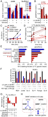
Glycine made by SHMT is required for B-lymphoma cell line growth. (A) Normalized total ion counts of nucleotide triphosphates in Jurkat ALL cells and Su-DHL-4 DLBCL cells after 72-h treatment with (+)-SHIN1 (5 µM). Coculture with 1 mM formate restores nucleotide levels selectively in Jurkat cells (mean ± SD, n = 3–6). (B) Normalized glutathione levels from Jurkat and Su-DHL-4 cells treated as in A (mean ± SD, n = 3–6). (C) Growth of Su-DHL-4 cells treated with (+)-SHIN1 and hypoxanthine (100 µM) or thymidine (16 µM; mean ± SD, n = 3). (D) Intracellular U-13C-glycine assimilation kinetics in Jurkat and Su-DHL-4 (gly, glycine; GSH, glutathione; mean ± SD, n = 3). (E) The steady-state labeling fraction of intracellular metabolites synthesized from glycine in cancer cell lines cultured in RPMI containing U-13C-glycine (mean ± SD, n = 3). (F) Cell growth (normalized to DMSO) of DLBCL and other hematopoietic cancer lines with 2.5 µM SHIN1, in RPMI with or without 1 mM formate and 10× physiological glycine (100 mg/L); all conditions included at least normal media glycine (10 mg/L; mean ± SD, n = 3). (G) Cell growth (or death) as measured by log2-fold change in cell number over 48 h in Su-DHL-4 cells cultured in RPMI with and without glycine (10 mg/L), formate (1 mM), the glycine transporter inhibitor RG1678 (300 nM), and/or (+)-SHIN1 (5 µM) (mean ± SD, n = 3). (H) Schematic illustrating the proposed glycine vulnerability in B cells. The SHMT reaction makes two products, 5,10-methylene–THF and glycine. When SHMT is inhibited, exogenous formate can be incorporated into the 1C cycle, whereas in B cells poor glycine uptake limits the ability of extracellular glycine to rescue.
Fig. S5.
Fig. S5.
SHMT inhibition in DLBCL cell lines results in depletion of glycine-derived metabolic products. (A) Normalized total ion counts of nucleotide triphosphates in Su-DHL-2 cells after 72 h treatment with (+)-SHIN1 (5 µM) and formate (1 mM; mean ± SD, n = 3). (B) Normalized total ion counts of nucleotide mono- and diphosphates in Jurkat and Su-DHL-4 cells after 72 h treatment with (+)-SHIN1 (5 µM) and formate (1 mM; mean ± SD, n = 3). (C) Formate (1 mM) rescues 1C shortage induced by SHIN1 (5 µM, 72 h) as evidenced by normalization of the levels of the purine intermediate AICAR (mean ± SD, n = 3). (D) Normalized glutathione levels from Jurkat and Su-DHL-2 cells treated as in A (mean ± SD, n = 3). (E) Normalized Su-DHL-4 growth after 48 h treatment with 2.5 µM (+)-SHIN1 and 250 µM glutathione (GSH; mean ± SD, n = 2). (F) Sensitivity of Su-DHL-4 cells to SHIN1 is dependent upon media glycine concentration. Cells were cultured in either normal RPMI (10 mg/L glycine) or RPMI containing 10× or 0.1× glycine and (+)-SHIN1 for 48 h, and growth was measured by resazurin assay (mean ± SD, n = 3). (G) Sensitivity of Jurkat cell growth to (+)-SHIN1 as a function of glycine concentration in RPMI in the same conditions as in E (mean ± SD, n ≥ 3).
Fig. S6.
Fig. S6.
SHIN1 treated Su-DHL-4 cells have sufficient glycine for tRNA charging. (A and B) Su-DHL-4 cells were treated with 5 µM (+)-SHIN1 or DMSO for 48 h before cell lysis, ribosome footprinting, and library purification. Abundance and phasing of sequenced reads shown. Highlighted reads (28–30 nt) were used for subsequent codon occupancy analysis. (C) Codon occupancy plotted against total codon frequency from ribosome profiling of Su-DHL-4 cells treated with DMSO (control). Shaded region is ±1 SD (σ) of the mean codon occupancy. Glycine codons (red) are highlighted. (D) Ratio of codon occupancies for SHIN1 treated Su-DHL-4 cells compared with DMSO control. Shaded region is ±1 SD (σ) of the mean ratio of codon occupancy. Glycine codons (red) are highlighted. Refer to SI Methods for details.

References

    1. Pavlova NN, Thompson CB. The emerging hallmarks of cancer metabolism. Cell Metab. 2016;23:27–47. - PMC - PubMed
    1. Schulze A, Harris AL. How cancer metabolism is tuned for proliferation and vulnerable to disruption. Nature. 2012;491:364–373. - PubMed
    1. Labuschagne CF, van den Broek NJF, Mackay GM, Vousden KH, Maddocks ODK. Serine, but not glycine, supports one-carbon metabolism and proliferation of cancer cells. Cell Rep. 2014;7:1248–1258. - PubMed
    1. Locasale JW, et al. Phosphoglycerate dehydrogenase diverts glycolytic flux and contributes to oncogenesis. Nat Genet. 2011;43:869–874. - PMC - PubMed
    1. Possemato R, et al. Functional genomics reveal that the serine synthesis pathway is essential in breast cancer. Nature. 2011;476:346–350. - PMC - PubMed

Publication types

MeSH terms

Associated data