. 2017 Oct 24;114(43):11476-11481.
doi: 10.1073/pnas.1709082114. Epub 2017 Oct 11.

FGF4 retrogene on CFA12 is responsible for chondrodystrophy and intervertebral disc disease in dogs

Affiliations

FGF4 retrogene on CFA12 is responsible for chondrodystrophy and intervertebral disc disease in dogs

Emily A Brown et al. Proc Natl Acad Sci U S A. .

Abstract

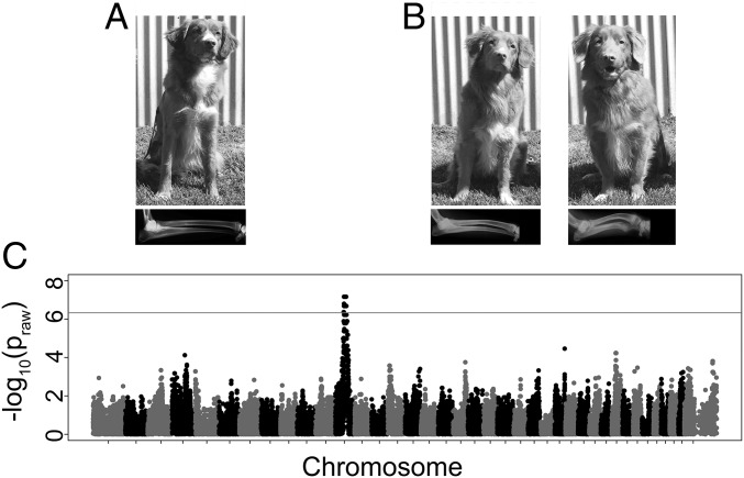
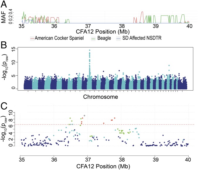
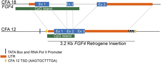
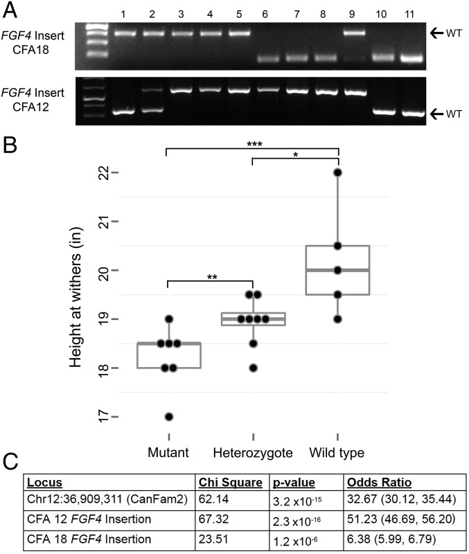
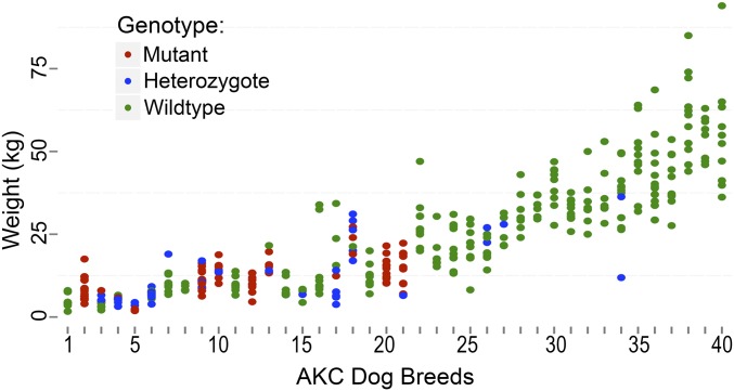
Chondrodystrophy in dogs is defined by dysplastic, shortened long bones and premature degeneration and calcification of intervertebral discs. Independent genome-wide association analyses for skeletal dysplasia (short limbs) within a single breed (PBonferroni = 0.01) and intervertebral disc disease (IVDD) across breeds (PBonferroni = 4.0 × 10⁻¹⁰) both identified a significant association to the same region on CFA12. Whole genome sequencing identified a highly expressed FGF4 retrogene within this shared region. The FGF4 retrogene segregated with limb length and had an odds ratio of 51.23 (95% CI = 46.69, 56.20) for IVDD. Long bone length in dogs is a unique example of multiple disease-causing retrocopies of the same parental gene in a mammalian species. FGF signaling abnormalities have been associated with skeletal dysplasia in humans, and our findings present opportunities for both selective elimination of a medically and financially devastating disease in dogs and further understanding of the ever-growing complexity of retrogene biology.

Keywords: GWAS; chondrodysplasia; dysplasia; genetic; inherited.


Conflict of interest statement

Conflict of interest statement: The University of California, Davis, has filed a provisional patent entitled: “Methods of Diagnosing Intervertebral Disc Disease and Chondrodystrophy in Canines,” on May 30, 2017.

Figures

Fig. 1.
Fig. 1.
Skeletal dysplasia in the NSDTR. (A) Picture and lateral thoracic limb radiograph of unaffected NSDTRs (ages 1 and 4 y old, respectively). (B, Left) Picture and lateral thoracic limb radiograph of mildly SD-affected NSDTRs (ages 4 and 2 y old, respectively); (Right) picture and lateral thoracic limb radiograph of more severely SD-affected NSDTRs (ages 3 y old and 6 mo old, respectively). Relative to the unaffected dog, the mildly SD-affected NSDTR has cranial bowing of the radius. Radiographic changes in the more severely SD-affected NSDTR include moderate cranial bowing of the radius, physeal widening, and incongruity of the elbow joint with the shape of the semilunar notch of the ulna being elongated. Pictures and radiographs are representatives of each phenotype and not paired. (C) SD in NSDTR GWAS. Manhattan plot showing −log10 of the raw P values for each genotyped SNP by chromosome (x axis). Genomic inflation was 1.02. Line denotes genome-wide significance based on Bonferroni-corrected P values.
Fig. 2.
Fig. 2.
Across breed investigation of SD-IVDD locus. (A) MAF on the y axis and base pair on CFA12 on the x axis plotted by breed. SD-affected NSDTR (n = 13), American cocker spaniel (n = 7), and beagle (n = 14). (B) Manhattan plot for the IVDD GWAS showing −log10 of the raw P values (y axis) for each genotyped SNP by chromosome (x axis). Genomic inflation was 1.63. (C) SNPs in 5-Mb region surrounding most highly associated SNP [chr12: 36,909,311 (canFam2)] plotted by base pair on the x axis and P value on the y axis. SNPs are color coded by r2 value to show the extent of linkage disequilibrium (orange, r2 0.6–0.8; green, r2 0.4–0.6; light blue, r2 0.2–0.4; and dark blue, r2 0–0.2).
Fig. 3.
Fig. 3.
Schematic of parental FGF4 (CFA18) retrotransposition to CFA12: 33,710,178 (canFam3): Predicted TATA box at chr12: 33,709,940–947 (canFam3) and predicted RNA Pol II promoter at chr12: 33,709,964–976 (canFam3) (54). Parental FGF4 UTRs are unknown in the dog; however, they were approximated based on human and mouse TransMap data (www.genome.ucsc.edu). The predicted 5′-UTR spans from chr18: 48,413,185 to 48,413,480 (canFam3), but RT-PCR in beagle IVD suggests that the TSS is between chr18: 48,413,315 and 48,413,402 (canFam3) (SI Appendix, SI Methods). The insert includes all predicted 3′-UTR, followed by 42 adenine residues and the duplicated TSD sequence [chr12: 33,710,168–33,710,178 (canFam3)].
Fig. 4.
Fig. 4.
Association of FGF4 insertion genotypes with height and IVDD. (A) Genotyping results for CFA18 and CFA12 FGF4 insertions. Arrows indicate wild-type (WT) band. Numbered lanes: ladder and 1–3, NSDTR; 4, beagle; 5, American cocker spaniel; 6, dachshund; 7, basset hound; 8, Pembroke Welsh corgi; 9, coton de Tulear; 10, cairn terrier; and 11, West Highland white terrier. (B) Height at the withers in inches (in) (y axis) by genotype (x axis) for 20 NSDTRs. Seven SD NSDTR cases were homozygous mutant for the CFA12 FGF4 insertion and their mean height was 18.22 in. Thirteen NSDTRs were unaffected with SD: 5 were wild type and had a mean height of 20.2 in; 8 were heterozygous for the CFA12 FGF4 insertion and had a mean height of 18.94 in. ***P = 0.01, **P = 0.02, *P = 0.03. (C) Association of loci with IVDD. The 95% confidence interval for odds ratio appears in parentheses.
Fig. 5.
Fig. 5.
CFA12 FGF4 genotypes across breeds: Genotypes for the CFA12 FGF4 insertion across dog breeds ordered by breed standard height from shortest to tallest (x axis), plotted by dog weight in kilograms (kg) (y axis). Dog breeds can be found in SI Appendix, Table S4. Only genotyped dogs with weights available (n = 376) are shown. Dogs are color coded by genotype status.

References

    1. Hansen H-J. A pathologic-anatomical study on disc degeneration in dog: With special reference to the so-called enchondrosis intervertebralis. Acta Orthop Scand. 1952;23(Suppl 11):1–130. - PubMed
    1. Hansen H-J. A pathologic-anatomical interpretation of disc degeneration in dogs. Acta Orthop Scand. 1951;20:280–293. - PubMed
    1. Braund KG, Ghosh P, Taylor TK, Larsen LH. Morphological studies of the canine intervertebral disc. The assignment of the beagle to the achondroplastic classification. Res Vet Sci. 1975;19:167–172. - PubMed
    1. Martinez S, Fajardo R, Valdes J, Ulloa-Arvizu R, Alonso R. Histopathologic study of long-bone growth plates confirms the basset hound as an osteochondrodysplastic breed. Can J Vet Res. 2007;71:66–69. - PMC - PubMed
    1. Bray JP, Burbidge HM. The canine intervertebral disk. Part two: Degenerative changes—Nonchondrodystrophoid versus chondrodystrophoid disks. J Am Anim Hosp Assoc. 1998;34:135–144. - PubMed

Publication types

MeSH terms

Substances

Supplementary concepts