. 2017 Oct 26;12(10):e0187077.
doi: 10.1371/journal.pone.0187077. eCollection 2017.

MiRNA-124 is a link between measles virus persistent infection and cell division of human neuroblastoma cells

Affiliations

MiRNA-124 is a link between measles virus persistent infection and cell division of human neuroblastoma cells

Hila Naaman et al. PLoS One. .

Abstract

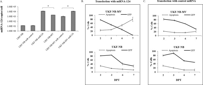
Measles virus (MV) infects a variety of lymphoid and non-lymphoid peripheral organs. However, in rare cases, the virus can persistently infect cells within the central nervous system. Although some of the factors that allow MV to persist are known, the contribution of host cell-encoded microRNAs (miRNA) have not been described. MiRNAs are a class of noncoding RNAs transcribed from genomes of all multicellular organisms and some viruses, which regulate gene expression in a sequence-specific manner. We have studied the contribution of host cell-encoded miRNAs to the establishment of MV persistent infection in human neuroblastoma cells. Persistent MV infection was accompanied by differences in the expression profile and levels of several host cell-encoded microRNAs as compared to uninfected cells. MV persistence infection of a human neuroblastoma cell line (UKF-NB-MV), exhibit high miRNA-124 expression, and reduced expression of cyclin dependent kinase 6 (CDK6), a known target of miRNA-124, resulting in slower cell division but not cell death. By contrast, acute MV infection of UKF-NB cells did not result in increased miRNA-124 levels or CDK6 reduction. Ectopic overexpression of miRNA-124 affected cell viability only in UKF-NB-MV cells, causing cell death; implying that miRNA-124 over expression can sensitize cells to death only in the presence of MV persistent infection. To determine if miRNA-124 directly contributes to the establishment of MV persistence, UKF-NB cells overexpressing miRNA-124 were acutely infected, resulting in establishment of persistently infected colonies. We propose that miRNA-124 triggers a CDK6-dependent decrease in cell proliferation, which facilitates the establishment of MV persistence in neuroblastoma cells. To our knowledge, this is the first report to describe the role of a specific miRNA in MV persistence.


Conflict of interest statement

Competing Interests: The authors have declared that no competing interests exist.

Figures

Fig 1
Fig 1. Characterization of UKF-NB-MV.
(A) Confocal images of fluorescent double staining of UKF-NB-MV cells with MV-P-protein antibodies (green); nuclear staining is demonstrated with DAPI (blue). Bars indicate original image sizes. (B) MV-P and M were detected by Western blot in UKF-NB-MV cells. (C) Hematoxylin-eosin staining of UKF-NB and UKFNB-MV. Both pictures were taken at the same magnification. (D) Time curve of cell growth and death. 2x10⁴ cells/well were seeded in 6 wells plates in triplicate. The average of cell proliferation and percentage of cell death were calculated from triplicates of three independent experiments. Averages with SEs of each point are indicated for three independent experiments, each point in triplicate. T-test was performed and significance of p<0.05 is shown (*). (E) Electron Microscopy of UKF-NB-MV cells. UKF-NB-MV cells were prepared for electron microscopy by conventional techniques. MV RNP (ribonucleoprotein) assembly particles (white arrow) and a rare instance of budding virus (black arrows) are shown in two magnifications. (F) UKF-NB-MV are more sensitive to cell death as compared to uninfected UKF-NB cells. 1.5x10⁵ cells/well UKF-NB-MV or UKF-NB cells were plated. 12 hours later, cisplatin was added at different concentrations in triplicate for 48 h, and the percentage of viable cells was determined by XTT. Experiments were repeated at least three times. The percent viability of each cell type is compared to untreated cells. Averages with SEs of each point are indicated for three independent experiments, each point in triplicate. T—test was performed and significance of p<0.05 is shown (*).
Fig 2
Fig 2. Differential expression of miRNAs in MV-persistently infected UKF-NB.
(A) Expression levels of selected miRNAs between uninfected and MV persistently infected UKF-NB cells. The relative expression (fold change) of each miRNA was determined by qPCR and expressed as the fold change relative to the control U6 gene. Expression in the UKF-NB cell line was used as the reference between the infected and the uninfected cells. Three independent cDNA libraries of each cell line were tested three times, each time point in triplicate. Averages with SEs are indicated for three independent experiments. (B) To determine the absolute transcript copy number per cell of miRNA-124 in UKF-NB-MV and uninfected UKF-NB cells (2X10⁵/well), we used miRNA-124 qSTAR primer pairs and a commercial miRNA-124 copy number standard kit. Averages with SEs are indicated for three independent experiments each in triplicate. T—test was performed and significance of at least p<0.05 is shown (*). (C) Relative expression of miRNA-124 in different clones of UKF-NB-MV cells. cDNA libraries of three independent UKF-NB-MV clones (I, II, III) were prepared and miRNA-124 relative expression was determined by qPCR and normalized to U6. Expression in the uninfected UKF-NB cell line was used as the reference. Triplicate samples of three independent cDNA libraries of each clone were tested. Clonal variations were observed, averages with SEs are indicated. (D) Analysis of qPCR of MV-P RNA in the different clones, normalized to U6. Expression in the uninfected UKF-NB cell line was used as the reference. Triplicate samples of three independent cDNA libraries of each clone were tested. Differences between all the clones were observed. UKF-NB-MV (fourth column) represents the parental un-cloned cells. Averages with SEs are indicated.
Fig 3
Fig 3. MiRNA-124 transfection induces apoptosis in UKF-NB-MV cells.
To study the possible effects of ectopic overexpression of miRNA-124 on UKF-NB and UKF-NB-MV cells and apoptosis, the cells were first transfected with GFP-miRNA-124 (miR124) or empty GFP-plasmid (miR control). (A) Three days post transfection (DPT) we determined the absolute transcript copy number per cell of miRNA-124. Following treatment, RNA was extracted, cDNA libraries were prepared, and miRNA-124 concentrations were determined using qSTAR primer pairs and a commercial miRNA-124 copy number standard kit. Averages with SEs are indicated for three independent experiments each in triplicate. T-test was performed and significance of at least p<0.05 is shown (*). (B) Apoptosis with Annexin V and 7AAD staining (early and late apoptosis respectively, added as a single value) in UKF-NB-MV and UKF-NB cells was determined following transfection (DPT) with GFP-miRNA-124 (miR124) or (C) control plasmid-GFP (miR control). Apoptosis was determined by flow cytometry exclusively in the GFP positive populations. Averages with SEs of four similar experiments in triplicate are indicated.
Fig 4
Fig 4. miRNA-124 overexpression induces cell death in UKF-NB-MV cells.
(A) UKF-NB-MV cells were transfected with: GFP-miRNA-124 (miR124), empty GFP-plasmid, or GFP-MUT-miRNA-124 (Mut124). The transfected cells were sorted 3 days post transfection (DPT) and seeded into six well plates. GFP fluorescence of viable cells was followed for 25 days post transfection. The experiment was repeated at least three times. Efficiency of transfection with the three plasmids was >90%. Inhibition of miRNA-124 expression by transfection with ANTAGOmiRNA-124 induces increased cell proliferation in UKF-NB-MV cells, but not in UKF-NB cells. (B) UKF-NB-MV and (C) UKF-NB cells were transfected with GFP-miRNA-124 (miR124), GFP-MUT-miRNA-124(Mut124), or negative control (NC), empty plasmid-GFP. Cell viability was determined by XTT assay at different times post transfection (hrs). Values were expressed as a percentage compared to the negative controls. Significance between samples was determined by T-test, and p<0.05 between samples is shown (*). Efficiency of transfection with the three plasmids was >90%. The experiment was repeated three times. (D) UKF-NB-MV and (E) UKF-NB cells were transfected with ANTAGO miRNA-124 (ANTAGO-miR124), ANTAGO scrambled miRNA-124 (ANTAGO scramble) or mock transfection reagents (N.C). Cell viability was determined by XTT at different times post transfection (hrs). The experiment was repeated three times. Significance between samples was determined by T-test, and p<0.05 between samples is shown (*). Efficiency of transfection with the three plasmids was >90%. (F) miRNA-124 silencing efficiency was tested on persistently infected UKF-NB-MV and persistently infected cell that were transfected with ANTAGO miRNA-124 or ANTAGO scrambled miRNA-124. MiRNAs relative expression was determined at 72 hrs by qPCR and expressed as the fold change relative to U6. Expression in the UKF-NB cell line was used as the reference. Three independent cDNA libraries of each cell line were tested three times each. Averages with SEs are indicated. Significance between samples was determined by T-test, and p<0.05 between samples is shown (*).
Fig 5
Fig 5. Downregulation of CDK6 following miRNA-124 expression.
(A) We isolated single cell clones from the UKF-NB-MV cell culture by limiting dilution. We then determined the expression level of CDK6 at three independent UKF-NB-MV clones (I, II, III). CDK6 was detected by Western blot. A representative experiment out of three is presented. Experiments were repeated at least three times and quantified by densitometry. Densitometry averages with SEs of three bands obtained from three independent experiments are indicated. (B) UKF-NB or UKF-NB-MV cells were transfected with miRNA-124- GFP (miR124), or GFP-MUT-miRNA-124(Mut124). GFP fluorescent cells were determined to be 95% in both groups two days post transfection. Expression of CDK6, MV-P protein and β-actin was determined by Western blot. Cell lysates were prepared three days post transfection. A representative experiment out of five is presented. Experiments were repeated five times and quantified by densitometry (lower panel). Densitometry averages with SEs of the bands obtained from five independent experiments are indicated. Significance between samples was determined by T-test, and p<0.05 between samples is shown (*). (C) Transfection of UKF-NB or UKF-NB-MV cells with ANTAGO miRNA-124 (ANTAGO-miR124), scrambled ANTAGO miRNA-124 (ANTAGO scramble), or transfection reagents alone (mock). Expression of CDK6, MV-P protein and β-actin was determined by Western blot. Cell lysates were prepared three days post transfection. Experiments were repeated five times and quantified by densitometry analysis (lower panel). Densitometry averages with SEs of the bands obtained from five independent experiments are indicated. Significance between samples was determined by T-test, and p<0.05 between samples is shown (*).
Fig 6
Fig 6. MiRNA-124 expression in MV acutely infected cells.
(A) UKF-NB cells were acutely infected with the MV Edmonston strain at various doses, and then assessed at either 3 or 7 DPI. (5x10³ PFU/ml, MOI 0.005(3 DPI), 5x10³ PFU/ml (7 DPI) or 5x10⁴ PFU/ml, MOI 0.05 (7 DPI)). Three or seven DPI, cDNA libraries were prepared and miRNA-124 relative expression was determined by qPCR and expressed as the fold change relative to U6. Expression in the uninfected UKF-NB cell line was used as a negative control, and persistently infected UKF-NB-MV was used as a positive control. Three independent cDNA libraries were tested three times in triplicate. Averages with SEs are indicated for three independent experiments. (B) Three DPI, cell lysates were prepared. MiRNA-124 target protein CDK6 and negative control (CDK4) were detected by Western blot in uninfected (UKF-NB) cells, persistently infected (UKF-NB-MV) and acute infected UKF-NB cells (5x10³ PFU/ml 3DPI). Experiments were repeated at least three times and quantified by densitometry analysis. A representative experiment is presented. Densitometry averages with SEs of three bands obtained from three independent experiments are indicated. Significance between samples was determined by T-test, and p<0.05 between samples is shown (*).
Fig 7
Fig 7. Stable miRNA-124 transfection of UKF-NB cells, followed by acute MV infection facilitated the establishment of persistently infected colonies.
2x10⁴ UKF-NB cells/well were seeded in triplicate. UKF-NB cells were stably transfected with constructs encoding GFP-miRNA-124 (miR124) or GFP-MUT-miRNA-124 (Mut124). Twenty days post transfection (DPT) GFP fluorescent cells were determined to be 95% in both groups. Cells monolayers were then acutely infected with Edmonston MV (5 x 10³ PFU /ml, MOI = 0.005). Most cells died, though surviving cells established colonies, which were scored. (A) Fixed and crystal-violet stained colonies were scored at day 7 and 21 post infection (DPI). The experiment was repeated three times. T-tests were performed and p<0.05 significance between samples is shown (*). (B) 21 DPI fluorescent staining of UKF-NB expressing GFP-miRNA-124(green), MV-P (red) and nuclear staining with DAPI (blue) is presented. Pictures were taken using confocal microscopy. All colonies were MV-positive and GFP-positive expressing the full length GFP-miRNA-124. (C) Establishment of MV persistent infection correlates with miRNA-124 expression. UKF-NB cells were seeded in triplicate. UKF-NB cells were stably transfected with constructs encoding GFP-miRNA-124(miR124) or the GFP-MUT-miRNA-124 (Mut124). Twenty days post transfection (DPT), GFP fluorescent cells were determined to be 95% in both groups. On day 21, cells monolayers were acutely infected with Edmonston MV. qPCR of miRNA-124 was determined before MV infection (0 DPI); 1 hour post infection (1 HPI) and 21 days post infection (DPI). The experiment was repeated three times for each independent infection. Averages with SEs are indicated. Significance between samples was determined by T-test, and p<0.05 between samples is shown (*).

Similar articles

Cited by

References

    1. Griffin DE, Lin WH, Pan CH. Measles virus, immune control, and persistence. FEMS Microbiol Rev. 2012;36(3):649–62. doi: 10.1111/j.1574-6976.2012.00330.x . - DOI - PMC - PubMed
    1. Moss WJ, Griffin DE. Measles Lancet. 379 England: 2012 Elsevier Ltd; 2012. p. 153–64. doi: 10.1016/S0140-6736(10)62352-5 - DOI - PubMed
    1. Lin WH, Kouyos RD, Adams RJ, Grenfell BT, Griffin DE. Prolonged persistence of measles virus RNA is characteristic of primary infection dynamics. Proc Natl Acad Sci U S A. 2012;109(37):14989–94. doi: 10.1073/pnas.1211138109 . - DOI - PMC - PubMed
    1. Schneider-Schaulies J, Niewiesk S, Schneider-Schaulies S, ter Meulen V. Measles virus in the CNS: the role of viral and host factors for the establishment and maintenance of a persistent infection. J Neurovirol. 1999;5(6):613–22. Epub 1999/12/22. . - PubMed
    1. Rall GF. Measles virus 1998–2002: progress and controversy. Annu Rev Microbiol. 2003;57:343–67. Epub 2003/10/07. doi: 10.1146/annurev.micro.57.030502.090843 . - DOI - PubMed