. 2017 Oct 26;12(10):e0186920.
doi: 10.1371/journal.pone.0186920. eCollection 2017.

Identification of novel substrates of Shigella T3SA through analysis of its virulence plasmid-encoded secretome

Affiliations

Identification of novel substrates of Shigella T3SA through analysis of its virulence plasmid-encoded secretome

Laurie Pinaud et al. PLoS One. .

Abstract

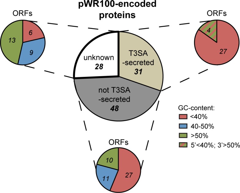
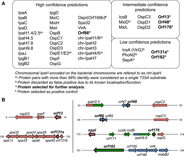
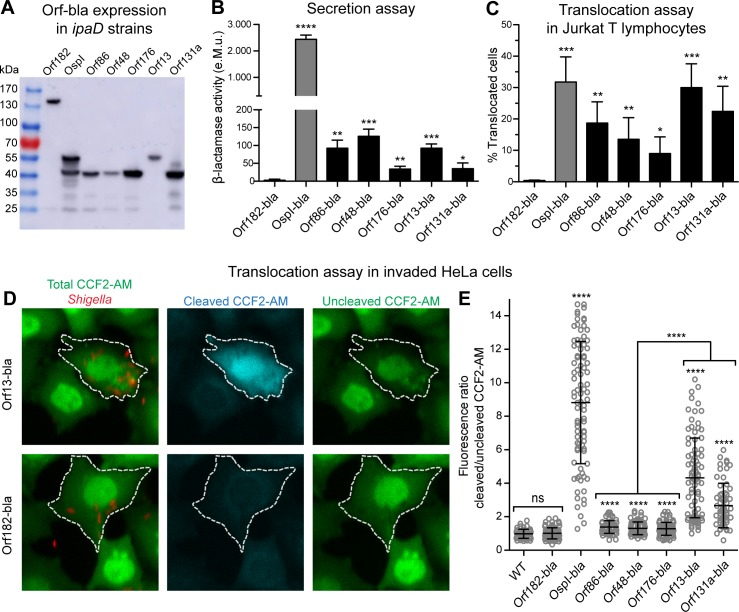
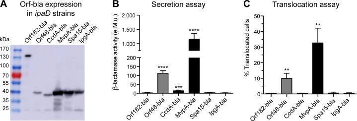
Many human Gram-negative bacterial pathogens express a Type Three Secretion Apparatus (T3SA), including among the most notorious Shigella spp., Salmonella enterica, Yersinia enterocolitica and enteropathogenic Escherichia coli (EPEC). These bacteria express on their surface multiple copies of the T3SA that mediate the delivery into host cells of specific protein substrates critical to pathogenesis. Shigella spp. are Gram-negative bacterial pathogens responsible for human bacillary dysentery. The effector function of several Shigella T3SA substrates has largely been studied but their potential cellular targets are far from having been comprehensively delineated. In addition, it is likely that some T3SA substrates have escaped scrutiny as yet. Indeed, sequencing of the virulence plasmid of Shigella flexneri has revealed numerous open reading frames with unknown functions that could encode additional T3SA substrates. Taking advantage of label-free mass spectrometry detection of proteins secreted by a constitutively secreting strain of S. flexneri, we identified five novel substrates of the T3SA. We further confirmed their secretion through the T3SA and translocation into host cells using β-lactamase assays. The coding sequences of two of these novel T3SA substrates (Orf13 and Orf131a) have a guanine-cytosine content comparable to those of T3SA components and effectors. The three other T3SA substrates identified (Orf48, Orf86 and Orf176) have significant homology with antitoxin moieties of type II Toxin-Antitoxin systems usually implicated in the maintenance of low copy plasmids. While Orf13 and Orf131a might constitute new virulence effectors contributing to S. flexneri pathogenicity, potential roles for the translocation into host cells of antitoxins or antitoxin-like proteins during Shigella infection are discussed.


Conflict of interest statement

Competing Interests: The authors have declared that no competing interests exist.

Figures

Fig 1
Fig 1. pWR100-encoded proteins.
Proteins encoded on pWR100 virulence plasmid classified based on their T3SA-dependent secretion and GC-content of the corresponding genes.
Fig 2
Fig 2. Mass spectrometry results.
(A) Group of proteins predicted to be T3SA substrates with low, medium and high confidence using the MS approach described in the text. Novel putative substrates and control substrate OspI selected for further analysis are highlighted in bold. (B) Genetic mapping of the ORFs selected for further investigation. Colours refer to the GC-content of the genes: red <40%; blue 40–50%; green >50%. Numbers refer to the coordinates on pWR100 sequence in kilobases (accession number AL391753).
Fig 3
Fig 3. Experimental confirmation of secretion and translocation of MS hits.
(A) ipaD Shigella lysates were analysed by immunoblotting with anti-β-lactamase antibody. Load equivalent to a bacterial culture optical density at 600 nm (OD600) of 0.2 for each lane. Chimeric proteins tested and their expected molecular weight (kiloDaltons, kDa): Orf182-bla (140), OspI-bla (53), Orf86-bla (40), Orf48-bla (41), Orf176-bla (40), Orf13-bla (51), Orf131a-bla (38). (B) Supernatants of ipaD strains expressing the Orf-bla chimeric proteins were incubated with nitrocefin. Enzymatic activity was calculated based on measurement of absorbance at 486 nm (A486). e.M.u.: equivalent Miller unit. Data are from 3 independent experiments. *p<0.05; **p<0.01; ***p<0.001; ****p<0.0001 (unpaired two-tailed Student’s t-test compared to Orf182-bla). (C) CCF2-AM-loaded Jurkat T lymphocytes were infected with WT strains expressing Orf-bla chimeric proteins for 1 hour. Data are from 4 independent experiments. Translocated cells were detected by flow cytometry. *p<0.05; **p<0.01; ***p<0.001 (unpaired two-tailed Student’s t-test compared to Orf182-bla). (D-E) CCF2-AM-loaded HeLa cells were infected with WT-DsRed strains expressing Orf-bla chimeric proteins for 1 hour, followed by 1 hour of incubation with gentamycin. (D) Representative images of cells infected with WT Shigella strains expressing the indicated chimeric proteins, showing total CCF2-AM (Green channel), cleaved CCF2-AM (Blue channel) and uncleaved CCF2-AM (FRET channel). Dashed lines denote cells invaded by DsRed bacteria (Red channel) that were used for fluorescence quantification. (E) CCF2-AM cleavage was quantified within invaded cells based on fluorescence intensity of cleaved over uncleaved CCF2-AM within the defined regions of interest. Data are from 3 independent experiments, representing 50 to 100 invaded cells analysed per condition. ****p<0.0001 (unpaired two-tailed Welch’s t-test compared to Orf182-bla unless depicted otherwise).
Fig 4
Fig 4. Investigation of secretion and translocation of antitoxins and chaperones.
(A) ipaD Shigella lysates were analysed by immunoblotting with anti-β-lactamase antibody. Load equivalent to a bacterial culture OD600 of 0.2 for each lane. Chimeric proteins tested and their expected molecular weight (kDa): Orf182-bla (140), Orf48-bla (41), CcdA-bla (38), MvpA-bla (38), Spa15-bla (44), IpgA-bla (44). (B) Supernatants of ipaD strains expressing the Orf-bla chimeric proteins were incubated with nitrocefin. Enzymatic activity was calculated based on measurement of A486nm. e.M.u.: equivalent Miller unit. Data are from 4 independent experiments. (C) CCF2-AM-loaded Jurkat T cells were infected with WT strains for 1 hour. Translocated cells were detected by flow cytometry. Data are from 3 independent experiments. (B-C) **p<0.01; ***p<0.001; ****p<0.0001 (unpaired two-tailed Student’s t-test compared to Orf182-bla).

References

    1. Galán JE, Lara-Tejero M, Marlovits TC, Wagner S. Bacterial type III secretion systems: specialized nanomachines for protein delivery into target cells. Annu Rev Microbiol. 2014;68: 415–438. doi: 10.1146/annurev-micro-092412-155725 - DOI - PMC - PubMed
    1. Ehsani S, Rodrigues CD, Enninga J. Turning on the spotlight—using light to monitor and characterize bacterial effector secretion and translocation. Curr Opin Microbiol. 2009;12: 24–30. doi: 10.1016/j.mib.2008.11.007 - DOI - PubMed
    1. Campbell-Valois FX, Sansonetti PJ. Tracking bacterial pathogens with genetically-encoded reporters. FEBS Lett. 2014;588: 2428–2436. doi: 10.1016/j.febslet.2014.05.022 - DOI - PubMed
    1. Sory MP, Boland A, Lambermont I, Cornelis GR. Identification of the YopE and YopH domains required for secretion and internalization into the cytosol of macrophages, using the cyaA gene fusion approach. Proc Natl Acad Sci USA. 1995;92: 11998–12002. - PMC - PubMed
    1. Charpentier X, Oswald E. Identification of the secretion and translocation domain of the enteropathogenic and enterohemorrhagic Escherichia coli effector Cif, using TEM-1 beta-lactamase as a new fluorescence-based reporter. J Bacteriol. 2004;186: 5486–5495. doi: 10.1128/JB.186.16.5486-5495.2004 - DOI - PMC - PubMed

Publication types