. 2017 Nov;5(6):1165-1173.
doi: 10.1111/andr.12415. Epub 2017 Oct 26.

Transdifferentiation of adult rat stem Leydig cells into prostatic and uterine epithelium, but not epidermis

Affiliations

Transdifferentiation of adult rat stem Leydig cells into prostatic and uterine epithelium, but not epidermis

M K Nanjappa et al. Andrology. 2017 Nov.

Abstract

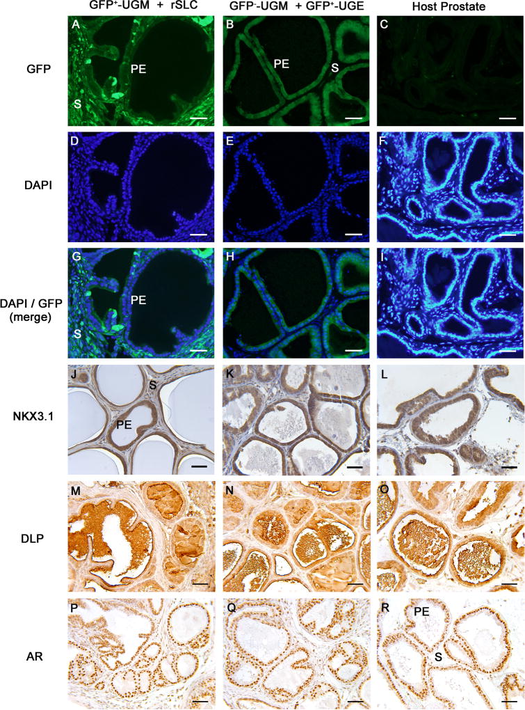
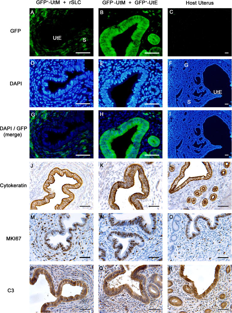
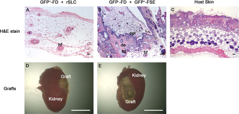
Stem Leydig cells (SLCs), precursors of testicular Leydig cells that secrete testosterone required for male sexual differentiation, spermatogenesis, and fertility, were recently identified in rat testes. Various types of stem cells have shown the ability to differentiate into other tissues, but there is no information on the plasticity of adult rat SLCs (rSLCs). This study investigated the ability of rSLCs to transdifferentiate into cell types from all three germ layers-prostatic epithelium (endoderm), uterine epithelium (mesoderm), and epidermis (ectoderm)-under the influence of inductive mesenchyme from fetal and neonatal tissues. To differentiate rSLCs into cells of other lineages, mesenchyme from green fluorescent protein (GFP)-expressing mice was used. Tissue recombinants of urogenital sinus mesenchyme (a potent prostate inducer) and rSLCs grafted into adult male hosts formed ductal structures resembling prostate after 5 weeks. Prostate epithelium was of rSLC origin as determined by absence of GFP expression, and expressed characteristic markers of prostatic epithelium. Similarly, uterine mesenchyme + rSLCs tissue recombinants contained a simple columnar epithelium that was histologically similar to normal uterine epithelium and expressed typical uterine epithelial markers, but was of rSLC origin. In contrast, epidermal tissue was absent in fetal dermis + rSLCs recombinants, suggesting rSLCs did not form skin epithelium. Thus, rSLCs can transdifferentiate into uterine and prostatic epithelium, mesodermal, and endodermal derivatives, respectively, but they may have a limited transdifferentiation potential, as shown by their inability to form epidermis, an ectodermal derivative.

Keywords: cell plasticity; ectoderm; endoderm; mesoderm; pluripotent; tissue recombination.


Figures

Figure 1
Figure 1. Rat stem Leydig cells (rSLCs) can transdifferentiate into prostatic epithelium (PE)
After 5 weeks of growth in vivo, GFP+-urogenital sinus mesenchyme (UGM) + rSLCs tissue recombinants developed into structures histologically resembling prostate and consisted of tubular structures lined by simple cuboidal epithelium supported by fibromuscular stroma (A, D, G, J, M, P). Critically, in GFP+-UGM + rSLCs grafts, the cuboidal epithelium (PE) was GFP, while the stroma (S) was GFP+ (A, G), indicating that the epithelium was of rSLC origin. This contrasts with control GFP-UGM + GFP+-urogenital sinus epithelium (UGE) tissue recombinants (B, E, H, K, N, Q) and host prostate (C, F, I, L, O, R), where GFP expression occurs only in epithelium (B, H) but not the stroma, or is seen in neither epithelium or stroma (C, I), respectively. In addition, the presumptive prostatic epithelium of rSLC origin robustly expressed a prostate epithelial-specific marker, NKX3.1 (J) that was also seen in GFP-UGM + GFP+-UGE tissue recombinants (K) and host prostate (L). In the epithelium of GFP+-UGM + rSLCs grafts, dorsolateral prostate secretory protein (DLP) production was seen in the epithelium and lumens of the ducts (M), while androgen receptor (AR) was expressed in all cells of these grafts (P). Both the secretory protein (DLP) expression and AR expression in these grafts was identical to control GFP-UGM + GFP+-UGE tissue recombinants (N, Q) and host prostate (O, R). Magnification bars in all panels are 50 µm. GFP, green fluorescent protein; DAPI, 4′,6-diamidino-2-phenylindole.
Figure 2
Figure 2. Rat stem Leydig cells (rSLCs) can transdifferentiate into uterine epithelium (UtE)
Tissue recombinants consisting of GFP+-uterine mesenchyme (UtM) + rSLCs (A, D, G, J, M, P) resembled control GFP-UtM + GFP+-UtE tissue recombinants (B, E, H, K, N, Q) that functioned as positive controls, as well as host uterus (C, F, I, L, O, R). Critically, in GFP+-UtM + rSLC grafts, the UtE was GFP (A, G), indicating rSLC origins. This contrasts with GFP-UtM + GFP+-UtE tissue recombinants and host uterus, where GFP is expressed only in epithelium (B, H), or not expressed (C, I), respectively. In addition, UtE derived from rSLCs expressed cytokeratin (J) and uterine secretory protein C3 (P), proteins that are typical of normal uterine epithelium, as shown by their expression in GFP-UtM + GFP+-UtE tissue recombinants (K, Q) and host uterus (L, R). Finally, GFP+-UtM + rSLC grafts (M) showed cell proliferation similar to GFP-UtM + GFP+-UtE tissue recombinants (N) and host uterus (O), as indicated by MKI67 immunostaining. Magnification bars of all panels are 50 µm. GFP, green fluorescent protein; DAPI, 4′,6-diamidino-2-phenylindole; S, stroma; G, glands.
Figure 3
Figure 3. Rat stem Leydig cells (rSLCs) do not transdifferentiate into epidermis
Tissue recombinants of GFP+-fetal dermis (FD) + rSLCs (A, D) produced small grafts that did not contain histologically identifiable skin and lacked an epidermis. In contrast, control GFP-FD + GFP+-fetal skin epidermis (FSE) tissue recombinants (B, E) produced large grafts of histologically normal skin containing keratinized epidermis (epi), dermis (de), sebaceous glands (sg) and hair follicles (hf) similar to host skin (C). Magnification bars are 50 µm for panels A–C and 5 mm for panels D and E.

Similar articles

Cited by

References

    1. Aboseif S, El-Sakka A, Young P, Cunha G. Mesenchymal reprogramming of adult human epithelial differentiation. Differentiation. 1999;65:113–118. - PubMed
    1. Benton L, Shan LX, Hardy MP. Differentiation of adult Leydig cells. J Steroid Biochem Mol Biol. 1995;53:61–68. - PubMed
    1. Boutin EL, Battle E, Cunha GR. The response of female urogenital tract epithelia to mesenchymal inductors is restricted by the germ layer origin of the epithelium: prostatic inductions. Differentiation. 1991;48:99–105. - PubMed
    1. Chen H, Jin S, Huang S, Folmer J, Liu J, Ge R, Zirkin BR. Transplantation of alginate-encapsulated seminiferous tubules and interstitial tissue into adult rats: Leydig stem cell differentiation in vivo? Mol Cell Endocrinol. 2016;436:250–258. - PMC - PubMed
    1. Cooke PS, Simon L, Nanjappa MK, Medrano TI, Berry SE. Plasticity of spermatogonial stem cells. Asian J Androl. 2015;17:355–359. - PMC - PubMed

LinkOut - more resources