Review
. 2017 Oct 26;20(11):1434-1447.
doi: 10.1038/nn.4661.

Viewpoints: how the hippocampus contributes to memory, navigation and cognition

Affiliations
Review

Viewpoints: how the hippocampus contributes to memory, navigation and cognition

John Lisman et al. Nat Neurosci. .

Erratum in

Abstract

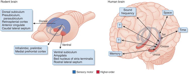
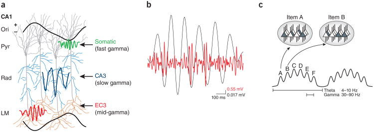
The hippocampus serves a critical function in memory, navigation, and cognition. Nature Neuroscience asked John Lisman to lead a group of researchers in a dialog on shared and distinct viewpoints on the hippocampus.

There has been a long history of studying the hippocampus, but recent work has made it possible to study the cellular and network basis of defined operations—operations that include cognitive processes that have been otherwise difficult to study (see Box 1 for useful terminology). These operations deal with the context-dependent representation of complex memories, the role of mental exploration based on imagined rather than real movements, and the use of recalled information for navigation and decision-making. The progress that has been made in understanding the hippocampus has motivated the study of other brain regions that provide hippocampal input or receive hippocampal output; the hippocampus is thus serving as a nucleating point for the larger goal of understanding the neural codes that allow inter-regional communication and more generally, understanding how memory-guided behavior is achieved by large scale integration of brain regions. In generating a discussion among experts in the study of the cognitive processes of the hippocampus, the editors and I have posed questions that probe important principles of hippocampal function. We hope that the resulting discussion will make clear to readers the progress that has been made, while also identifying issues where consensus has not yet been achieved and that should be pursued in future research. – John Lisman


Figures

Figure 1
Figure 1
Homologous regions of the hippocampus in the human and rat brains. The ventral quadrant of the rodent hippocampus became disproportionally enlarged in primates to keep up with the increasingly larger share of higher-order neocortex and formed the uncus and body. Only the relatively small tail part of the primate hippocampus communicates with visuospatial areas. This tail is the part that is homologous with the rodent dorsal-intermediate hippocampus. Differential connections to and from the different segments of the septotemporal axis are shown. Most recordings and manipulations in the rodent brain have been performed in the dorsal hippocampus. Adapted with permission from ref. 145, “Distinct representations and theta dynamics in dorsal and ventral hippocampus”
Figure 2
Figure 2
Comparison of global remapping of hippocampal place cells to rate remapping. Place cell firing rate is plotted as a function of rat position along linear track. (a) Global remapping occurs in response to a context shift resulting from use of different tracks or the same track but with different behavioral relevance. Inset at right shows that the average position of place cell peak firing in one map is not correlated with peak position in the new map. (b) Rate remapping occurs following minor changes in context and produces changes in place cell firing rates but no change in the position of maximal activity. Inset at right shows that the average position of peak place cell firing in original map is highly correlated with peak position in the new map.
Figure 3
Figure 3
Hippocampal pyramidal neurons provide organized responses to spatial stimuli, nonspatial stimuli, and time. (a) Top: in a simple navigation task, a rat runs back and forth along a linear track. Bottom: place cells, each represented by a different color, are activated as the rat runs along the track. Adapted with permission from ref. 146, Nature Publishing Group. (b) Top: rats alternated between left and right paths on a T-maze separated by running on a wheel. Bottom: time cells, each represented by a different color, are activated as time elapses during wheel running. Adapted with permission from ref. 82, Nature Publishing Group. (c) Top: in the sound-manipulation task depicted in a, rats learned to press and hold a joystick to increase the frequency of a pure tone until it reached a target frequency range, and to then release the joystick in order to obtain a reward. Bottom: CA1 place cells were active around particular frequencies, which resulted in sequences of firing fields that tiled the auditory frequency space. Their activity scaled with trial duration, contracting in fast trials (left) and expanding in slow trials (right). Similar results were obtained with grid cells. These results demonstrate that cells in the hippocampal formation can represent the gradually increasing frequency of a tone in a map-like fashion, like they do for other continuous dimensions such as space and time. Adapted with permission from ref. 68, Nature Publishing Group.
Figure 4
Figure 4
Properties and utilization of grid cells. (a) Top left: grid-cell network in the MEC showing hexagonally spaced bumps of activity (red, peak rate). Either a true velocity signal or an imagined velocity signal is put into the grid-cell network. Top right: the bumps move leftward in the network in proportion to a true velocity vector derived from optic flow information, somatosensory information or vestibular information, thus performing an integration process. Cells in the network shown and those in similar networks but with different grid-cell spacing converge onto hippocampal cells and generate place cells (having maximal activity at a given position; bottom left. The position of active place cells moves left as result of the movement of activity bumps in the grid cell networks (bottom right). Adapted with permission from ref. 98, Elsevier. (b) Path integration occurs during a complex path taken by the animal, resulting in hippocampal place cell activity at a position that is directly relatable to the actual position of the animal even though no sensory information about the place was used in the process. Inset: one mechanism for finding a straight path to the rat's home is to generate an imagined velocity vector and put it into the grid cell network. This moves active place cells along the line of the arrow shown. If this activity leads to the excitation of ‘home’ place cells, the direction of the vector is the straightest direction of motion to get the rat home.
Figure 5
Figure 5
Properties of theta and gamma oscillations in the hippocampus. (a) Dendritic layer gamma oscillations report activity of upstream neurons. Entorhinal layer 3 (EC3) mid-frequency gamma input (60–100 Hz) modulates distal dendrites in stratum lacunosum moleculare (LM) at the positive peak of CA1 pyramidal layer theta (Pyr), followed by CA3-projected slow-gamma (30–60 Hz) input in stratum radiatum (Rad) on the descending theta phase. The gamma power ratios and their relative timing determine the theta phase of spiking of CA1 pyramidal cells. High-frequency (90–150 Hz) gamma power at the trough of the theta cycle (not shown) largely reflects spiking activity. Ori, stratum oriens. Adapted with permission from ref. 131, Elsevier. (b) Simultaneous filtered (20–100 Hz) intracellular recording from a CA1 pyramidal neuron (red) and extracellular recording (black) from the CA1 pyramidal layer. (c) Neural code organized by theta and gamma oscillations. Ovals at the top represent the shifting states of the network during two gamma cycles (spiking cells are dark gray and constitute the ensemble that codes for a particular item). Each gamma cycle contains a unique constellation of neurons (ensembles A to F). Adapted with permission from ref. 147, Oxford Academic Journals.

References

    1. Tulving E, Donaldson W, Bower GH. Organization of Memory. Academic Press; 1972.
    1. O'Keefe J, Nadel L. The Hippocampus as a Cognitive Map. Clarendon Press; Oxford University Press; 1978.
    1. Cohen NJ, Eichenbaum H. Memory, Amnesia, and the Hippocampal System. MIT Press; 1993.
    1. Deuker L, Bellmund JL, Navarro Schröder T, Doeller CF. eLife. 2016;5:e16534. - PMC - PubMed
    1. Nielson DM, Smith TA, Sreekumar V, Dennis S, Sederberg PB. Proc Natl Acad Sci USA. 2015;112:11078–11083. - PMC - PubMed

Publication types