Ultra high-field SWI of the substantia nigra at 7T: reliability and consistency of the swallow-tail sign
- PMID: 29073886
- PMCID: PMC5658950
- DOI: 10.1186/s12883-017-0975-2
Ultra high-field SWI of the substantia nigra at 7T: reliability and consistency of the swallow-tail sign
Abstract
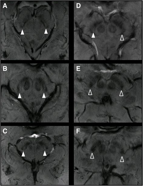
Background: The loss of the swallow-tail sign of the substantia nigra has been proposed for diagnosis of Parkinson's disease. Aim was to evaluate, if the sign occurs consistently in healthy subjects and if it can be reliably detected with high-resolution 7T susceptibility weighted imaging (SWI).
Methods: Thirteen healthy adults received SWI at 7T. 3 neuroradiologists, who were blinded to patients' diagnosis, independently classified subjects regarding the swallow-tail sign to be present or absent. Accuracy, positive and negative predictive values (PPV and NPV) as well as inter- and intra-rater reliability and internal consistency were analyzed.
Results: The sign could be detected in 81% of the cases in consensus reading. Accuracy to detect the sign compared to the consensus was 100, 77 and 96% for the three readers with PPV reader 1/2/3 = 1/0.45/0.83 and NPV = 1/1/1. Inter-rater reliability was excellent (inter-class correlation coefficient = 0.844, alpha = 0.871). Intra-rater reliability was good to excellent (reader 1 R/L = 0.625/0.786; reader 2 = 0.7/0.64; reader 3 = 0.9/1).
Conclusion: The swallow-tail sign can be reliably detected. However, our data suggest its occurrence is not consistent in healthy subjects. It may be possible that one reason is an individually variable molecular organization of nigrosome 1 so that it does not return a uniform signal in SWI.
Keywords: 7 tesla; Nigrosome 1; Parkinson’s disease; SWI; Swallow-tail sign; Ultra high-field MRI.
Conflict of interest statement
Ethics approval and consent to participate
The Clinical Investigation Ethics Committee of the University of Erlangen-Nuremberg approved the protocol of this prospective study and the research was conducted in accordance with the Declaration of Helsinki. All participants gave written informed consent prior to all measurements and agreed upon publication.
Consent for publication
Not applicable.
Competing interests
The authors declare that they have no competing interests.
Publisher’s Note
Springer Nature remains neutral with regard to jurisdictional claims in published maps and institutional affiliations.
Figures


Similar articles
-
The 'swallow tail' appearance of the healthy nigrosome - a new accurate test of Parkinson's disease: a case-control and retrospective cross-sectional MRI study at 3T.PLoS One. 2014 Apr 7;9(4):e93814. doi: 10.1371/journal.pone.0093814. eCollection 2014. PLoS One. 2014. PMID: 24710392 Free PMC article.
-
Microvessels may Confound the "Swallow Tail Sign" in Normal Aged Midbrains: A Postmortem 7 T SW-MRI Study.J Neuroimaging. 2019 Jan;29(1):65-69. doi: 10.1111/jon.12576. Epub 2018 Nov 8. J Neuroimaging. 2019. PMID: 30407679
-
Using 'swallow-tail' sign and putaminal hypointensity as biomarkers to distinguish multiple system atrophy from idiopathic Parkinson's disease: A susceptibility-weighted imaging study.Eur Radiol. 2017 Aug;27(8):3174-3180. doi: 10.1007/s00330-017-4743-x. Epub 2017 Jan 19. Eur Radiol. 2017. PMID: 28105503
-
7 Tesla magnetic resonance imaging: a closer look at substantia nigra anatomy in Parkinson's disease.Mov Disord. 2014 Nov;29(13):1574-81. doi: 10.1002/mds.26043. Epub 2014 Oct 12. Mov Disord. 2014. PMID: 25308960 Review.
-
Nigrosome 1 imaging: technical considerations and clinical applications.Br J Radiol. 2019 Sep;92(1101):20180842. doi: 10.1259/bjr.20180842. Epub 2019 Jun 5. Br J Radiol. 2019. PMID: 31067082 Free PMC article. Review.
Cited by
-
Parkinsonism in the psychiatric setting: an update on clinical differentiation and management.BMJ Neurol Open. 2020 Jan 27;2(1):e000034. doi: 10.1136/bmjno-2019-000034. eCollection 2020. BMJ Neurol Open. 2020. PMID: 33681781 Free PMC article. Review.
-
Dorsal hyperintensity and iron deposition patterns in the substantia nigra of Parkinson's disease, idiopathic REM sleep behavior disorder, and Parkinson-plus syndromes at 7T MRI: a prospective diagnostic study.Transl Neurodegener. 2025 Jul 4;14(1):35. doi: 10.1186/s40035-025-00495-4. Transl Neurodegener. 2025. PMID: 40616129 Free PMC article.
-
Substantia Nigra Radiomics Feature Extraction of Parkinson's Disease Based on Magnitude Images of Susceptibility-Weighted Imaging.Front Neurosci. 2021 May 31;15:646617. doi: 10.3389/fnins.2021.646617. eCollection 2021. Front Neurosci. 2021. PMID: 34135726 Free PMC article.
-
Imaging the Nigrosome 1 in the substantia nigra using susceptibility weighted imaging and quantitative susceptibility mapping: An application to Parkinson's disease.Neuroimage Clin. 2020;25:102103. doi: 10.1016/j.nicl.2019.102103. Epub 2019 Nov 20. Neuroimage Clin. 2020. PMID: 31869769 Free PMC article.
-
Studying Alzheimer disease, Parkinson disease, and amyotrophic lateral sclerosis with 7-T magnetic resonance.Eur Radiol Exp. 2021 Aug 26;5(1):36. doi: 10.1186/s41747-021-00221-5. Eur Radiol Exp. 2021. PMID: 34435242 Free PMC article. Review.
References
-
- Schwarz ST, Abaei M, Gontu V, Morgan PS, Bajaj N, Auer DP. Diffusion tensor imaging of nigral degeneration in Parkinson's disease: a region-of-interest and voxel-based study at 3 T and systematic review with meta-analysis. NeuroImage Clin. 2013;3:481–488. doi: 10.1016/j.nicl.2013.10.006. - DOI - PMC - PubMed
MeSH terms
LinkOut - more resources
Full Text Sources
Other Literature Sources
Medical

