. 2017 Dec:75:184-188.
doi: 10.1016/j.oraloncology.2017.10.013. Epub 2017 Nov 1.

Improving margin revision: Characterization of tumor bed margins in early oral tongue cancer

Affiliations

Improving margin revision: Characterization of tumor bed margins in early oral tongue cancer

Arpan V Prabhu et al. Oral Oncol. 2017 Dec.

Abstract

Objectives: To improve margin revision, this study characterizes the number, fragmentation, and orientation of tumor bed margins (TBM) in patients with pT1-2 pN0 squamous cell carcinoma (SCC) of the oral tongue.

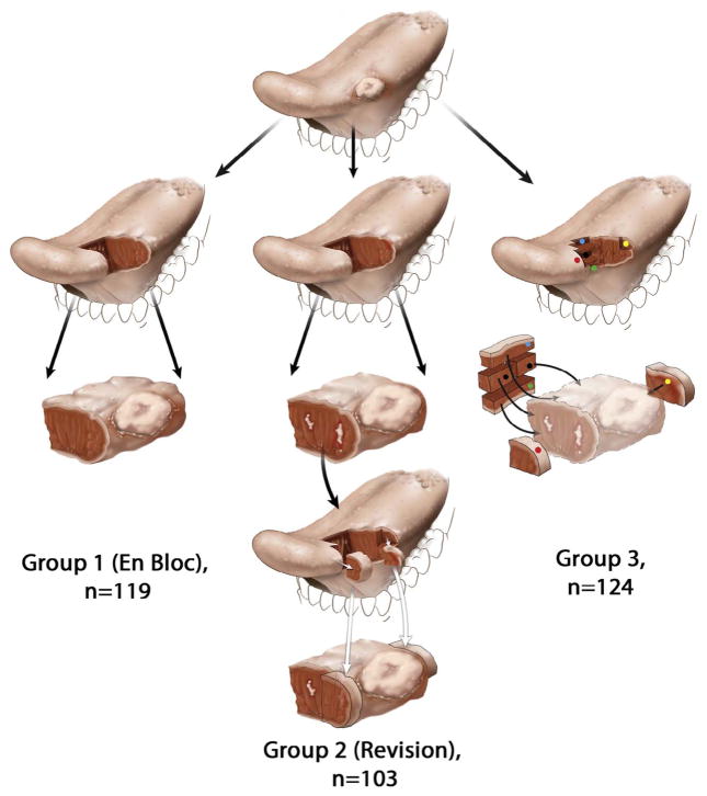
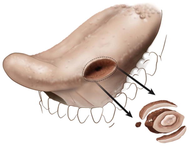
Materials and methods: Pathology reports (n=346) were reviewed. TBM parameters were indexed. In Group 1 patients all margins were obtained from the glossectomy specimen and there were no TBM. In Revision Group/Group 2 (n=103), tumor bed was sampled to revise suboptimal margins identified by examination of the glossectomy specimen. In Group 3 (n=124), TBM were obtained before examination of the glossectomy specimen.

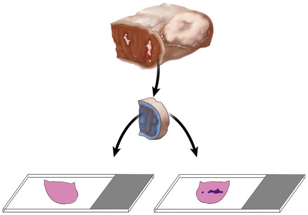
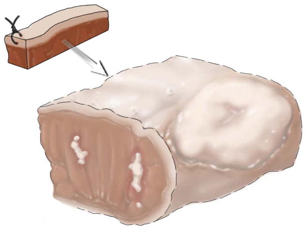
Results and conclusions: Fewer TBMs were obtained per patient in Group 2 compared to Group 3 (57/103, 55% of patients with <3 vs. 117/124, 94%, ≥3 TBMs, respectively). The new margin surface was more frequently indicated in Group 2 compared to Group 3 (59/103, 57%, vs. 19/124, 15%, p<.001). If glossectomy specimen margins are accepted as the reference standard, then the TBM was 15% sensitive in Group 2 (95% confidence interval [CI], 7-29) and 32% sensitive in Group 3 (95% CI, 15-55). TBM fragmentation (23/103, 22% vs. 42/124, 34%) and frozen vs. permanent discrepancies (8/103, 3% vs. 3/124, 2%) were similar between Groups 2 and 3. The new margin surface was not indicated in 6 of 11 cases with discrepant frozen vs. permanent pathology findings, precluding judgment on final margin status. To facilitate the assessment of final margins, TBM should be represented by one tissue fragment with a marked new margin surface.

Keywords: Margin fragmentation; Margin orientation; Oral tongue; Revision; Specimen margin; Squamous cell carcinoma; Tumor bed margin.


Conflict of interest statement

Conflict of Interest and Disclosure

None.

Figures

Fig. 1
Fig. 1
Schematic representation of the glossectomy workflow. For simplicity, a predominantly exophytic tumor at the lateral edge of the oral tongue is illustrated, with the row representing the next step in the workflow. In Group 1 (left column), tumor bed margins were not sampled. In Group 2 (middle column), margins were examined from the glossectomy specimen and found to be positive or otherwise suboptimal. The surgeon revised margins by obtaining additional tissue from the tumor bed. White irregular areas in the anterior aspect of glossectomy specimen represent residual carcinoma at the initial anterior margin (third row). The surgeon revised anterior margin by obtaining additional tissue from the tumor bed, representing a new anterior margin (fourth row). To imagine the relationship between the actual glossectomy margins and additional tissue, the two types of margins are superimposed in the fifth row. Due to the challenges of relocating the exact aspect of the relevant anterior margin in the tumor bed, size discrepancy, and uncertain orientation of the additional tissue, it is conceivable that in some cases the revised margin may not actually cover the entire residual tumor at the anterior glossectomy margin. In Group 3 (right column), five margins are primarily sampled from the tumor bed (red, green, yellow, blue, and black dots), without prior examination of the glossectomy specimen (displayed in lighter colors in the third row) by the pathologist.
Fig. 2
Fig. 2
Approach to margin assessment and number of tumor bed margins per patient. For more than half of Group 2 patients (57/103, 55%) < 3 tumor bed margins were taken. In Group 3, ≥3 tumor bed margins were obtained in all but 7 patients (117/124, 94%).
Fig. 3
Fig. 3
An illustration of how tumor bed margins are likely sampled for smaller tumors (partial glossectomy with 3 tumor bed margins).
Fig. 4
Fig. 4
Unequivocal orientation of the revised tumor bed margin. The new margin surface of the revised anterior margin from left partial glossectomy is indicated by ink. The white irregular area in the anterior aspect of glossectomy specimen represents residual carcinoma at the initial anterior margin. The dark blue irregular area on the right glass slide represents tumor. Without orientation, the surface to be first examined intraoperatively is picked randomly. When indicated, the new margin surface will be examined first intraoperatively. If the frozen section is negative for tumor, but the permanent section of the frozen remnant reveals tumor, the overall margin status is “close, but negative”. Such determination is impossible if the tumor bed margin was fragmented or the new true margin surface was not indicated. In addition to ink, new margin surface can be indicated by a stitch, clip, or directly by surgeon. This revision margin is thin and is best processed as shave margin. Thicker margins can be processed as radial margins (new margin surface re-inked, cut into, and embedded on edge).
Fig. 5
Fig. 5
Indirect orientation of the revised tumor bed margin. The anterior aspect of the new margin is indicated by stitch. However, if the relationship between the tumor bed margin and main resection specimen is unknown, the new margin surface may be difficult to figure out.

References

    1. Weinstock YE, Alava I, 3rd, Dierks EJ. Pitfalls in determining head and neck surgical margins. Oral Maxillofac Surg Clin North Am. 2014;26:151–62. - PubMed
    1. Chen AY. Quality initiatives in head and neck cancer. Curr Oncol Rep. 2010;12:109–14. - PubMed
    1. Black C, Marotti J, Zarovnaya E, Paydarfar J. Critical evaluation of frozen section margins in head and neck cancer resections. Cancer. 2006;107:2792–800. - PubMed
    1. Meier JD, Oliver DA, Varvares MA. Surgical margin determination in head and neck oncology: current clinical practice. The results of an International American Head and Neck Society Member Survey. Head Neck. 2005;27:952–8. - PubMed
    1. Hinni ML, Ferlito A, Brandwein-Gensler MS, Takes RP, Silver CE, Westra WH, et al. Surgical margins in head and neck cancer: a contemporary review. Head Neck. 2013;35:1362–70. - PubMed

Publication types