. 2018 Mar 1;314(3):L493-L504.
doi: 10.1152/ajplung.00372.2017. Epub 2017 Oct 26.

Autophagy and the unfolded protein response promote profibrotic effects of TGF-β1 in human lung fibroblasts

Affiliations

Autophagy and the unfolded protein response promote profibrotic effects of TGF-β1 in human lung fibroblasts

Saeid Ghavami et al. Am J Physiol Lung Cell Mol Physiol. .

Abstract

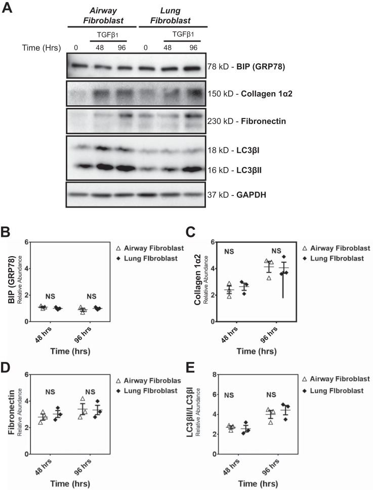
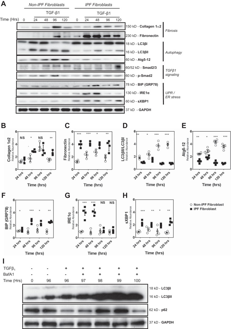
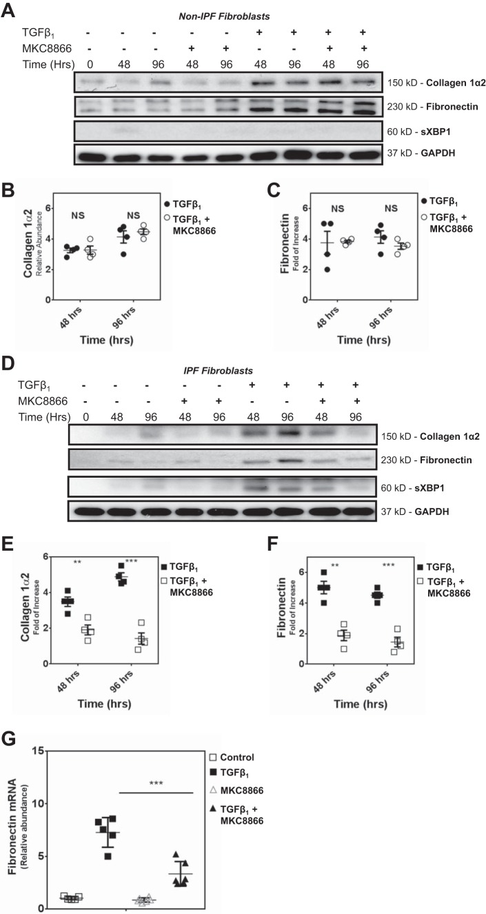
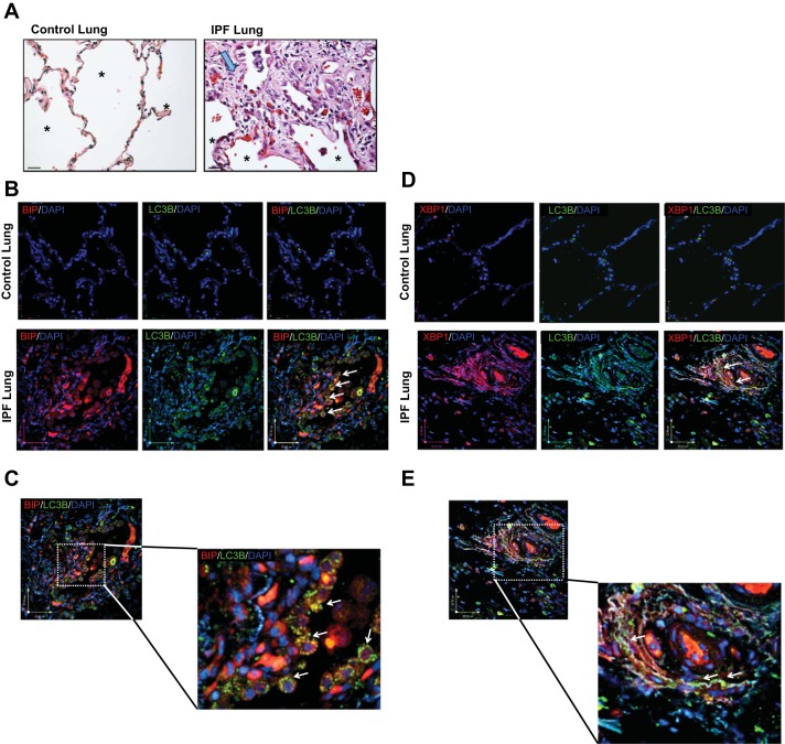
Idiopathic pulmonary fibrosis (IPF) is a lethal fibrotic lung disease in adults with limited treatment options. Autophagy and the unfolded protein response (UPR), fundamental processes induced by cell stress, are dysregulated in lung fibroblasts and epithelial cells from humans with IPF. Human primary cultured lung parenchymal and airway fibroblasts from non-IPF and IPF donors were stimulated with transforming growth factor-β1 (TGF-β1) with or without inhibitors of autophagy or UPR (IRE1 inhibitor). Using immunoblotting, we monitored temporal changes in abundance of protein markers of autophagy (LC3βII and Atg5-12), UPR (BIP, IRE1α, and cleaved XBP1), and fibrosis (collagen 1α2 and fibronectin). Using fluorescent immunohistochemistry, we profiled autophagy (LC3βII) and UPR (BIP and XBP1) markers in human non-IPF and IPF lung tissue. TGF-β1-induced collagen 1α2 and fibronectin protein production was significantly higher in IPF lung fibroblasts compared with lung and airway fibroblasts from non-IPF donors. TGF-β1 induced the accumulation of LC3βII in parallel with collagen 1α2 and fibronectin, but autophagy marker content was significantly lower in lung fibroblasts from IPF subjects. TGF-β1-induced collagen and fibronectin biosynthesis was significantly reduced by inhibiting autophagy flux in fibroblasts from the lungs of non-IPF and IPF donors. Conversely, only in lung fibroblasts from IPF donors did TGF-β1 induce UPR markers. Treatment with an IRE1 inhibitor decreased TGF-β1-induced collagen 1α2 and fibronectin biosynthesis in IPF lung fibroblasts but not those from non-IPF donors. The IRE1 arm of the UPR response is uniquely induced by TGF-β1 in lung fibroblasts from human IPF donors and is required for excessive biosynthesis of collagen and fibronectin in these cells.

Keywords: IRE1; pulmonary fibrosis; spliced XBP1; transforming growth factor-β1.


Figures

Fig. 1.
Fig. 1.
Human airway and lung fibroblasts isolated from nonidiopathic pulmonary fibrosis (non-IPF) lungs do not differ in transforming growth factor-β1 (TGF-β1)-induced fibrotic response, unfolded protein response (UPR), and autophagy activity. A: primary human non-IPF airway and lung fibroblasts (passages 3–6) were treated with TGF-β1 (2.5 ng/ml) (0–96 h). Cell lysates were collected and the abundance of UPR (BIP), autophagy (LC3βII), and profibrotic proteins (collagen 1α2 and fibronectin) were assessed by Western blot. Protein loading was confirmed using GAPDH. AE are representative of experiments performed on 3 different primary non-IPF airway and lung fibroblast cultures (n = 3). No differences (P > 0.05) were detected in UPR (B), fibrotic (C and D), and autophagy (E) markers between airway and lung fibroblasts. BE: dot plots of data from individual experiments. The horizontal line in each column represents the mean, with error bars showing SE. NS, no significant difference.
Fig. 2.
Fig. 2.
Fibroblasts isolated from non-IPF lungs and those with IPF donors differ in TGF-β1-induced fibrotic response, UPR, and autophagy activity. A: primary human non-IPF fibroblast and IPF fibroblasts (passages 3–6) were treated with TGF-β1 (2.5 ng/ml) (0–120 h). Cell lysates were collected and subjected to SDS-PAGE, and the expression of UPR [BIP, IRE1α, and sliced XBP1 (sXBP1)], autophagy (LC3βII and Atg5-12), and extracellular matrix (ECM) proteins (collagen 1α2 and fibronectin), as well as Smad signaling effectors were assessed by Western blot analysis. Protein loading was confirmed using GAPDH. Data in A represent experiments performed on 4 different primary non-IPF and IPF fibroblasts. B and C: densitometry showed that TGF-β1 stimulation led to an increase in abundance of collagen Iα2 and fibronectin that was greater in IPF fibroblasts compared with non-IPF fibroblasts. D and E: abundance of autophagy proteins LC3βII/LC3βI and Atg5-12 following TGF-β1 stimulation was significantly lower in IPF fibroblasts as compared with non-IPF fibroblasts. FH: TGF-β1 stimulation also resulted in significant accumulation of UPR proteins (BIP, IRE1α, and sXBP), which was greater in IPF fibroblasts compared with non-IPF fibroblasts. Data in BH are shown in dot plots of from 4 individual experiments using different IPF and non-IPF samples. The horizontal line in each column represents the mean ± SE. ***P < 0.001; **P < 0.01: *P < 0.05; NSP > 0.05, no significant difference. I: primary human IPF fibroblasts (passages 2–3) were treated with TGF-β1 (2.5 ng/ml) for 96 h and then treated with bafiloECM A1 (Baf-A1; 100 nM) for up to 4 h more. LC3βII accumulation and p62 degradation were measured using GAPDH as protein loading confirmed.
Fig. 3.
Fig. 3.
Autophagy is required for TGF-β1-induced fibrosis in both non-IPF and IPF fibroblasts. A: primary human non-IPF fibroblasts were treated with TGF-β1 (2.5 ng/ml) in the presence or absence of the chemical inhibitors of autophagy (Baf-A1; 10 nM) for the indicated times. A representative blot from 4 different primary non-IPF fibroblasts (n = 4) is shown. B and C: densitometry analysis of different IPF and non-IPF fibroblasts confirmed that the inhibition of autophagy significantly inhibited TGF-β1-induced collagen biosynthesis in human non-IPF fibroblasts while there is not any significant difference in LC3βII between TGF-β1 and TGF-β1 + Baf-A1 group. Densitometry data are normalized to time matched controls grown in insulin/transferrin/selenium medium. Data are shown in dot plots of from 4 individual experiments using different cell lines. The horizontal line in each column represents the mean, with error bars showing SE. ***P < 0.001; NSP > 0.05, no significant difference. D: primary human IPF fibroblasts were treated with TGF-β1 (2.5 ng/ml) in the presence or absence of the autophagy chemical inhibitor bafiloECM A1 (10 nM) for the indicated time points. Immunoblots show the expression of collagen 1α2 and LC3βII. Data in D represent examples of 4 different primary IPF fibroblasts (n = 4). E and F: densitometry analysis confirmed that autophagy inhibition significantly inhibited TGF-β1-induced collagen biosynthesis in human IPF fibroblasts while there is not any significant difference in LC3βII between TGF-β1 and TGF-β1 + Baf-A1 group. Densitometry data are normalized to time-matched controls in ITS medium. Data are shown in dot plots of from 4 individual experiments using different cell lines. The horizontal line in each column represents the mean, with error bars showing SE. *** P < 0.001; NSP > 0.05. no significant difference.
Fig. 4.
Fig. 4.
XBP splicing is required for TGF-β1-induced fibrosis in primary human IPF, but not non-IPF, fibroblasts. A: primary human non-IPF fibroblasts were stimulated with TGF-β1 (2.5 ng/ml) in the presence or absence of the IRE1α inhibitor MKC8866 (10 μM) for the indicated time points. The representative immunoblot shows the abundance of collagen 1α2, fibronectin, and sXBP in 4 different primary non-IPF fibroblasts. B and C: densitometry analysis (n = 4) confirmed that MKC8866 did not significantly reduce TGF-β1-induced collagen 1α2 or fibronectin biosynthesis in non-IPF human fibroblasts. Data are shown in dot plots of from 4 individual experiments using different cell lines. The horizontal line in each column represents the mean, with error bars showing SE. NSP > 0.05, no significant difference. D: primary human IPF fibroblasts were stimulated with TGF-β1 (2.5 ng/ml) in the presence or absence of IRE1 inhibitor (10 μM) for the indicated time points. Immunoblots are arranged to show expression of collagen 1α2, fibronectin and sXBP. The immunoblot is representative of experiments performed on 4 different primary IPF fibroblasts (n = 4). E and F: densitometry analysis confirmed that treatment of IPF fibroblasts with IRE1 inhibitor significantly reduced TGF-β1-induced collagen 1α2 and fibronectin biosynthesis. Data are shown in dot plots of from 4 individual experiments using different cell lines. The horizontal line in each column represents the mean, with error bars showing SE. ***P < 0.001; **P < 0.01. G: primary human IPF fibroblasts were stimulated with TGF-β1 (2.5 ng/ml) in the presence or absence of IRE1 inhibitor (10 μM) for 96 h. For a baseline control, cells were maintained in serum- and TGF-β1-deficient media. Quantitative PCR showed that MKC8866 significantly inhibited TGF-β1-induced fibronectin mRNA accumulation in IPF fibroblasts. The experiments were performed in duplicate on 2 different primary IPF fibroblast cell lines. Data are shown in dot plots from each experiment. The horizontal line in each column represents the mean, with error bars showing SE. ***P < 0.001.
Fig. 5.
Fig. 5.
Autophagy flux inhibition and IRE1α inhibition modulates UPR and autophagy in IPF fibroblasts. Primary human IPF fibroblasts were stimulated with TGF-β1 (2.5 ng/ml) in the presence or absence of bafilomycin-A1 (10 nM) or IRE1 inhibitor MKC8866 (10 μM) for 48 or 96 h. Immunoblots show abundance of autophagy (LC3βII, p62) and UPR (BIP) markers. The blot shown is representative of experiments performed on 2 different primary IPF fibroblast cell lines.
Fig. 6.
Fig. 6.
Autophagy and UPR proteins colocalize and are simultaneously increased in IPF lungs. A: representative histological images of peripheral parenchymal non-IPF and IPF lung tissue stained with hematoxylin and eosin. IPF lung tissue showing fibroblastic foci (large arrow). Alveolar structures are indicated by an asterisk. Magnification: ×40. B and C: immunofluorescence confocal microscopy of control and IPF lung tissue for LC3βII (green), BIP (red), and DAPI (blue). Punctate foci of LC3βII are indicated with white arrows. D and E: confocal images of control and IPF lung tissue for LC3βII (green), XBP (red), and DAPI (blue). Scale bars are included in each image.

Similar articles

Cited by

References

    1. Adamson YI, Bowden DH. Pulmonary injury and repair. Organ culture studies of murine lung after oxygen. Arch Pathol Lab Med 100: 640–643, 1976. - PubMed
    1. Ahluwalia N, Shea BS, Tager AM. New therapeutic targets in idiopathic pulmonary fibrosis. Aiming to rein in runaway wound-healing responses. Am J Respir Crit Care Med 190: 867–878, 2014. doi:10.1164/rccm.201403-0509PP. - DOI - PMC - PubMed
    1. Araya J, Kojima J, Takasaka N, Ito S, Fujii S, Hara H, Yanagisawa H, Kobayashi K, Tsurushige C, Kawaishi M, Kamiya N, Hirano J, Odaka M, Morikawa T, Nishimura SL, Kawabata Y, Hano H, Nakayama K, Kuwano K. Insufficient autophagy in idiopathic pulmonary fibrosis. Am J Physiol Lung Cell Mol Physiol 304: L56–L69, 2013. doi:10.1152/ajplung.00213.2012. - DOI - PubMed
    1. Borges FT, Melo SA, Özdemir BC, Kato N, Revuelta I, Miller CA, Gattone VH II, LeBleu VS, Kalluri R. TGF-β1-containing exosomes from injured epithelial cells activate fibroblasts to initiate tissue regenerative responses and fibrosis. J Am Soc Nephrol 24: 385–392, 2013. doi:10.1681/ASN.2012101031. - DOI - PMC - PubMed
    1. Budinger GR, Mutlu GM, Eisenbart J, Fuller AC, Bellmeyer AA, Baker CM, Wilson M, Ridge K, Barrett TA, Lee VY, Chandel NS. Proapoptotic Bid is required for pulmonary fibrosis. Proc Natl Acad Sci USA 103: 4604–4609, 2006. doi:10.1073/pnas.0507604103. - DOI - PMC - PubMed

Publication types

MeSH terms

LinkOut - more resources