Review
. 2017 Nov;18(11):1905-1921.
doi: 10.15252/embr.201643426. Epub 2017 Oct 26.

The ins and outs of endoplasmic reticulum-controlled lipid biosynthesis

Affiliations
Review

The ins and outs of endoplasmic reticulum-controlled lipid biosynthesis

Julie Jacquemyn et al. EMBO Rep. 2017 Nov.

Abstract

Endoplasmic reticulum (ER)-localized enzymes synthesize the vast majority of cellular lipids. The ER therefore has a major influence on cellular lipid biomass and balances the production of different lipid categories, classes, and species. Signals from outside and inside the cell are directed to ER-localized enzymes, and lipid enzyme activities are defined by the integration of internal, homeostatic, and external information. This allows ER-localized lipid synthesis to provide the cell with membrane lipids for growth, proliferation, and differentiation-based changes in morphology and structure, and to maintain membrane homeostasis across the cell. ER enzymes also respond to physiological signals to drive carbohydrates and nutritionally derived lipids into energy-storing triglycerides. In this review, we highlight some key regulatory mechanisms that control ER-localized enzyme activities in animal cells. We also discuss how they act in concert to maintain cellular lipid homeostasis, as well as how their dysregulation contributes to human disease.

Keywords: SREBP; mTOR; CCTα; de novo lipid synthesis; lipin.


Figures

Figure 1
Figure 1. The organization of ER‐localized GPL/GL synthesis
(A) The GPL/GL enzyme network where membrane lipids are in purple, storage lipids are in orange, and water‐soluble intermediates are in blue. Only key nodes are shown. The enzymes shown in gray are transmembrane ER proteins, while blue indicates a protein that reversibly associates with the ER membrane. Double arrow highlights the feedback loop where PtdA activates CDP–choline synthesis, which is a key control point for PtdCho production. Dotted lines indicate multiple reactions, and “*” indicates that PEMT has a restricted expression pattern in animals and its physiological importance is largely reported for hepatocytes. (B) Lipid synthesis organization within the ER system. Schematic of ER organization in a simple animal cell. There are structurally and/or functionally distinct domains, such as the inner and outer nuclear membrane (INM and ONM), and specialized contact sites between the ER and most organelles (not all shown). The localization of lipid enzymes within the ER system is still poorly defined, but some new concepts are emerging including the following: (I) Enzymes of the TAG pathway can relocalize from a broad distribution to concentrate at sites of growing lipid droplets. (II) Enzymes of PtdSer synthesis (PSS1 and PSS2) concentrate at mitochondria‐associated membranes that are also the sites where newly synthesized PtdSer is transferred to mitochondria. (III) The lipin and CCTα enzymes that reversibly bind membranes also carry NLS and shuttle between the cytosol and nucleus through nuclear pore complexes. This affects whether these enzymes interact with the main‐ER membrane or inner nuclear membrane. The LBR sterol reductase is also a well‐characterized INM resident. AGPAT, 1‐acylglycerol‐3‐phosphate O‐acyltransferase; CEPT, choline/ethanolamine phosphotransferase; CK, choline kinase; CDP, cytidine diphosphate; Cho, choline; CDS, PtdA cytidylyltransferase; EPT, ethanolamine phosphotransferase; G‐3‐P, glycerol 3‐phosphate; PtdGly, phosphatidylglycerol; PSS, phosphatidylserine synthase; MAM, mitochondria‐associated membrane.
Figure 2
Figure 2. Mechanisms of ER membrane homeostasis
(A) Hepatocyte membrane compositions. The ER membranes are enriched in GPL and contain little sphingolipid or cholesterol. PtdA is also detected in the ER membrane, but not the plasma membrane. PtdIns includes phosphorylated and non‐phosphorylated species. These data are modified from 39 and are in line with several other studies, although not all lipid species were assessed in each report 120, 133, 134. (B) Intrinsic properties of lipids effect membrane properties. PtdCho is a cylindrical lipid where the headgroup and acyl chains occupy a similar lateral area, while PtdEtn is conical because it has a smaller headgroup. PtdCho spontaneously assembles into bilayers and stabilizes bilayers in vivo, while PtdEtn promotes voids between lipid headgroups within a bilayer and forms curved “hexagonal” structures in isolation 135. Note that the looser packing associated with PtdEtn also exposes small lipid headgroups, such as that of PtdA, for protein interactions 136. Acyl chain saturation also affects membrane properties such as thickness and lipid packing density 137, 138. (C) CCTα is activated by membrane absorption of the M‐domain that is regulated by lipid packing and PtdA. Activated CCTα produces the CDP–choline that is typically rate‐limiting for PtdCho production and therefore membrane biogenesis. (D) Immature, inactive SREBP is a transmembrane ER protein. It is activated when altered membrane environment causes SCAP to change conformation and dissociate from Insig. This alters SREBP/SCAP trafficking (not shown) and allows protease‐dependent release of the mature SREBP transcription factor from the membrane. This then can enter the nucleus and activate gene transcription via binding to SRE, including genes of lipid synthesis. (E) The IRE1/XBP1(S) branch of the UPR is activated by increased lipid saturation and packing density in the ER membrane. This causes IRE1 dimerization, self‐phosphorylation of cytosolic residues, and activation of IRE1 RNA splicing activity that removes an intron from the XBP1 mRNA. The XBP1(S) protein is then translated and acts as a transcription factor. (F) Lipin PtdA‐phosphatase activity requires that it integrates into a membrane. This is positively regulated by PtdA and negatively regulated by lipin phosphorylation state. Lipin activity often promotes TAG production while reducing PtdCho. Pi, orthophosphate.
Figure 3
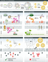
Figure 3. The regulation of ER‐localized lipid synthesis in cell growth, differentiation, and disease
(A) SREBP‐1 and/or XBP1(S) signaling are important for cellular adaptation of lipid synthesis to cell growth, differentiation (like secretory pathway expansion), or the environment (like TAG production in response to nutrients). The mechanisms that allow context‐dependent variation in the final outcome of these pathways remain under study. (B) Growth signals, including key pathways such as insulin signaling, stimulate increased lipid synthesis associated with increased cell biomass. The activation of cell growth‐associated lipid synthesis typically requires mTORC1 and couples to SREBP‐1 including enhancing SREBP‐1 cleavage. Pro‐growth signaling also consistently causes lipin phosphorylation, which converts this enzyme from a nuclear localization to a cytosolic localization, and negatively correlates with PtdA‐phosphatase activity. This in turn disinhibits CCTα by reducing PtdA metabolism to DAG. Lipin PtdA‐phosphatase activity also inhibits SREBP by sequestering cleaved SREBP at the nuclear periphery. The inner nuclear membrane CTDNEP/NEP1R1 phosphatase complex maintains dephosphorylated lipin and thus counters pro‐growth lipin phosphorylation 112, 138. Pi, orthophosphate. (C) Pathological impact of elevated TAG production in hepatocytes caused by excess nutrients. These cells are functionally specialized to convert carbohydrates into TAG. High‐level TAG production from excess nutrients alters the ER membrane lipid saturation profile and the ratio between PtdCho and PtdEtn. This altered membrane lipid composition induces broader ER dysfunction, included impairing calcium transport and protein misfolding.

References

    1. Carman GM, Han GS (2011) Regulation of phospholipid synthesis in the yeast Saccharomyces cerevisiae . Annu Rev Biochem 80: 859–883 - PMC - PubMed
    1. Fernandez‐Murray JP, McMaster CR (2016) Lipid synthesis and membrane contact sites: a crossroads for cellular physiology. J Lipid Res 57: 1789–1805 - PMC - PubMed
    1. Henry SA, Kohlwein SD, Carman GM (2012) Metabolism and regulation of glycerolipids in the yeast Saccharomyces cerevisiae . Genetics 190: 317–349 - PMC - PubMed
    1. Shindou H, Hishikawa D, Harayama T, Eto M, Shimizu T (2013) Generation of membrane diversity by lysophospholipid acyltransferases. J Biochem 154: 21–28 - PubMed
    1. Harayama T, Eto M, Shindou H, Kita Y, Otsubo E, Hishikawa D, Ishii S, Sakimura K, Mishina M, Shimizu T (2014) Lysophospholipid acyltransferases mediate phosphatidylcholine diversification to achieve the physical properties required in vivo . Cell Metab 20: 295–305 - PubMed

Publication types

MeSH terms

Substances