

Roles of miRNAs and long noncoding RNAs in the progression of diabetic retinopathy
- PMID: 29074557
- PMCID: PMC5705777
- DOI: 10.1042/BSR20171157
Roles of miRNAs and long noncoding RNAs in the progression of diabetic retinopathy
Abstract
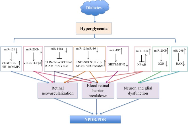
Diabetic retinopathy (DR) is the leading cause of blindness in working-age adults across the world. The pathogenesis of DR is multifactorial and the molecular mechanisms are still not fully understood. Accumulating evidence has demonstrated that noncoding RNAs (ncRNAs) may be aberrantly expressed and may play vital roles in the development of DR. Amongst ncRNAs, miRNAs and long ncRNAs (lncRNAs) are known for their regulatory functions. Here, we summarize the functions and mechanisms of known aberrantly expressed miRNAs and lncRNAs in DR. Additionally, a novel lncRNA-mRNA-miRNA network is included in this review. We highlight original studies that provide detailed data about the mechanisms of miRNAs and lncRNAs, their applications as diagnostic or prognostic biomarkers, and their potential therapeutic targets. In conclusion, this review will help us gain a better understanding of the molecular mechanisms by which miRNAs and lncRNAs perform their functions in DR, and provide general strategies and directions for future research.
Keywords: diabetic retinopathy; dysregulation; long noncoding RNA; microRNA; molecular mechanisms.
© 2017 The Author(s).
Conflict of interest statement
The authors declare that there are no competing interests associated with the manuscript.
Figures
Similar articles
-
MicroRNA and diabetic retinopathy-biomarkers and novel therapeutics.Ann Transl Med. 2021 Aug;9(15):1280. doi: 10.21037/atm-20-5189. Ann Transl Med. 2021. PMID: 34532417 Free PMC article. Review.
-
miRNA, lncRNA and circRNA: Targeted Molecules Full of Therapeutic Prospects in the Development of Diabetic Retinopathy.Front Endocrinol (Lausanne). 2021 Nov 10;12:771552. doi: 10.3389/fendo.2021.771552. eCollection 2021. Front Endocrinol (Lausanne). 2021. PMID: 34858342 Free PMC article. Review.
-
Aberrant expression of long noncoding RNAs in early diabetic retinopathy.Invest Ophthalmol Vis Sci. 2014 Feb 18;55(2):941-51. doi: 10.1167/iovs.13-13221. Invest Ophthalmol Vis Sci. 2014. PMID: 24436191
-
A comprehensive competitive endogenous RNA network pinpoints key molecules in diabetic retinopathy.Mol Med Rep. 2019 Feb;19(2):851-860. doi: 10.3892/mmr.2018.9715. Epub 2018 Dec 3. Mol Med Rep. 2019. PMID: 30535492 Free PMC article.
-
Mechanistic and therapeutic perspectives of non-coding RNA-modulated apoptotic signaling in diabetic retinopathy.Cell Biol Toxicol. 2024 Jul 6;40(1):53. doi: 10.1007/s10565-024-09896-z. Cell Biol Toxicol. 2024. PMID: 38970639 Free PMC article. Review.
Cited by
-
Malignancy-related mir-210, mir-373 and let-7 levels are affected in iron deficiency anemia.Afr Health Sci. 2023 Sep;23(3):245-253. doi: 10.4314/ahs.v23i3.30. Afr Health Sci. 2023. PMID: 38357103 Free PMC article.
-
Dyslipidemia in retinal metabolic disorders.EMBO Mol Med. 2019 Oct;11(10):e10473. doi: 10.15252/emmm.201910473. Epub 2019 Sep 5. EMBO Mol Med. 2019. PMID: 31486227 Free PMC article. Review.
-
Differential expression analysis of mRNAs, lncRNAs, and miRNAs expression profiles and construction of ceRNA networks in PEDV infection.BMC Genomics. 2022 Aug 13;23(1):586. doi: 10.1186/s12864-022-08805-0. BMC Genomics. 2022. PMID: 35964002 Free PMC article.
-
Long non-coding RNA VIM Antisense RNA 1 (VIM-AS1) sponges microRNA-29 to participate in diabetic retinopathy.Acta Diabetol. 2020 Sep;57(9):1111-1116. doi: 10.1007/s00592-020-01536-2. Epub 2020 May 23. Acta Diabetol. 2020. PMID: 32447557 Free PMC article.
-
MicroRNA and diabetic retinopathy-biomarkers and novel therapeutics.Ann Transl Med. 2021 Aug;9(15):1280. doi: 10.21037/atm-20-5189. Ann Transl Med. 2021. PMID: 34532417 Free PMC article. Review.
References
-
- Schwartz S.S., Epstein S., Corkey B.E., Grant S.F.A., Gavin J.R. Iii, Aguilar R.B. et al. (2017) A unified pathophysiological construct of diabetes and its complications. Trends Endocrinol. Metab. 28, 645–655 - PubMed
-
- Antonetti D.A., Klein R. and Gardner T.W. (2012) Diabetic retinopathy. N. Engl. J. Med. 366, 1227–1239 - PubMed
-
- Ciulla T.A., Amador A.G. and Zinman B. (2003) Diabetic retinopathy and diabetic macular edema: pathophysiology, screening, and novel therapies. Diabetes Care 26, 2653–2664 - PubMed
-
- Frank R.N. (2004) Diabetic retinopathy. N. Engl. J. Med. 350, 48–58 - PubMed
Publication types
MeSH terms
Substances
LinkOut - more resources
Full Text Sources
Other Literature Sources
Medical