Review
. 2018 Jan 29;131(2):jcs203331.
doi: 10.1242/jcs.203331.

Phragmoplast microtubule dynamics - a game of zones

Affiliations
Review

Phragmoplast microtubule dynamics - a game of zones

Andrei Smertenko et al. J Cell Sci. .

Abstract

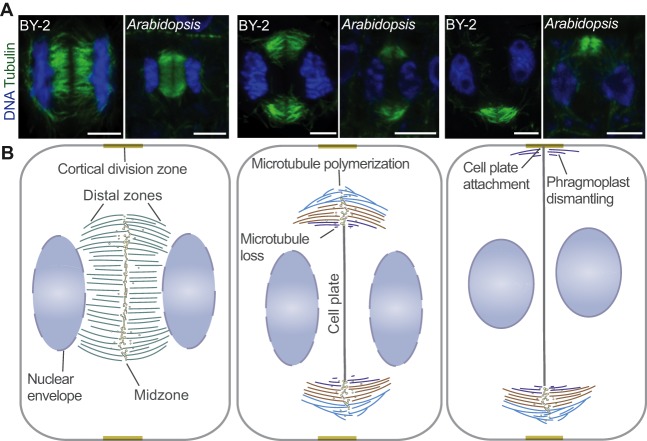
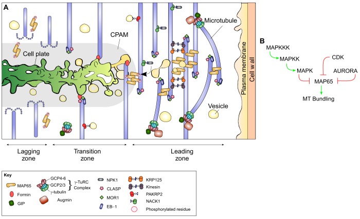
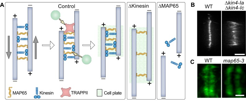
Plant morphogenesis relies on the accurate positioning of the partition (cell plate) between dividing cells during cytokinesis. The cell plate is synthetized by a specialized structure called the phragmoplast, which consists of microtubules, actin filaments, membrane compartments and associated proteins. The phragmoplast forms between daughter nuclei during the transition from anaphase to telophase. As cells are commonly larger than the originally formed phragmoplast, the construction of the cell plate requires phragmoplast expansion. This expansion depends on microtubule polymerization at the phragmoplast forefront (leading zone) and loss at the back (lagging zone). Leading and lagging zones sandwich the 'transition' zone. A population of stable microtubules in the transition zone facilitates transport of building materials to the midzone where the cell plate assembly takes place. Whereas microtubules undergo dynamic instability in all zones, the overall balance appears to be shifted towards depolymerization in the lagging zone. Polymerization of microtubules behind the lagging zone has not been reported to date, suggesting that microtubule loss there is irreversible. In this Review, we discuss: (1) the regulation of microtubule dynamics in the phragmoplast zones during expansion; (2) mechanisms of the midzone establishment and initiation of cell plate biogenesis; and (3) signaling in the phragmoplast.

Keywords: Cell plate; Cytokinesis; Microtubule dynamics; Phragmoplast.


Conflict of interest statement

Competing interestsThe authors declare no competing or financial interests.

Figures

Fig. 1.
Fig. 1.
Successive stages of phragmoplast expansion. (A) Representative pictures of successive stages of phragmoplast expansion in Tobacco BY-2 and Arabidopsis root meristem cells. In both systems, the phragmoplast expands asymmetrically and first attaches to one side of the mother cell, where microtubules depolymerize. Microtubules were immunostained with anti-tubulin antibody (green) and staining of DNA with DAPI (blue). Scale bars: 5 µm. (B) The phragmoplast is established between daughter nuclei (blue ellipses) during the anaphase-to-telophase transition and at this stage is disk shaped. Cell plate assembly takes place in the midzone. Once the cell plate assembly reaches the tubular network stage, microtubules depolymerize and this results in a ring-shaped phragmoplast. The ring phragmoplast then expands towards the cortical division zone, which was established during prophase by the preprophase band. During the expansion, microtubules depolymerize in the regions where the cell plate reaches a certain degree of maturity (lagging zone, purple) and polymerize at the outermost phragmoplast edge (leading zone, light blue). The leading and lagging zones sandwich transition zone (orange). Phragmoplast expansion is generally asymmetric and the cell plate first fuses with one side of the cortical division zone (Cutler and Ehrhardt, 2002; Lucas and Sack, 2012). The phragmoplast is dismantled once the cell plate attaches to the plasma membrane and the cell wall.
Fig. 2.
Fig. 2.
Regulation of microtubule processes in the phragmoplast zones. (A) The key events in the phragmoplast zones. In the leading zone, branching and cytoplasmic microtubules are generated by the γ-TuRC-mediated nucleation. Polymerization of microtubules is supported by a family of plus-end-binding proteins. Anti-parallel microtubules are bundled by MAP65 (orange). This overlap of anti-parallel microtubules defines the midzone and recruits nascent cell plate vesicles (pale yellow). In the transition zone, microtubules undergo nucleation, polymerization and depolymerization. Microtubules may be stabilized through formin-mediated contacts with the nascent cell plate membrane or interaction with yet unknown components of the CPAM. Kinesins transport cell plate materials and MAPKKK NPK1 along microtubules. Cell plate vesicles undergo fusion and tubulation, resulting in a tubulo-vesicular network (pale yellow). Callose accumulates in the cell plate (green gradient). In the lagging zone, microtubule-stabilizing proteins become inactivated through phosphorylation and microtubules depolymerize. (B) Multiple kinase pathways regulate the interaction between MAP65 and microtubules. The MAPK cascade, CDKs and Aurora kinases phosphorylate different residues on the C-terminal domain of MAP65. Phosphorylation weakens the affinity of MAP65 for microtubules and inhibits its ability to bundle anti-parallel microtubules. Green lines indicate activation and red lines indicate inhibition.
Fig. 3.
Fig. 3.
Mechanics of the phragmoplast midzone establishment. (A) MAP65 (orange) bundles anti-parallel microtubules (pale blue) in the midzone. Anterograde kinesins (blue) bind to anti-parallel microtubules and slide them apart in opposite directions, as shown by gray arrows. Eventually, the thickness of the midzone is determined by the balance of forces generated by kinesin movements and the affinity of MAP65 for microtubules. MAP65 interacts with the tethering complex TRAPPII. Tethering complexes can initiate cell plate assembly by recruiting vesicles (green circles) to the midzone. In the absence of kinesins (ΔKinesin), both the midzone and cell plate are wider. In the absence of MAP65 (ΔMAP65), kinesins slide microtubules away from each other. Consequently, cell plate formation is abrogated. (B) Example picture of the localization of MAP65a-citrine in Physcomytrella caulonemal tip cell. Reprinted from de Keijzer et al., 2017, with permission from Elsevier. Double knockout of Kin4-Ia and Kin4-Ic results in wider microtubule overlap as evident by MAP65a–citrine localization in living cells. (C) Example picture of how loss of MAP65-3 activity (Arabidopsis ple-6 allele) leads to a wider phragmoplast midzone. Reprinted from Muller et al., 2004, with permission from Elsevier. Microtubules were immunostained with anti-tubulin antibody (green). WT, wild type. Scale bars: 5 μm (B), 2 µm (C).

Similar articles

Cited by

References

    1. Aidemark M., Andersson C.-J., Rasmusson A. G. and Widell S. (2009). Regulation of callose synthase activity in situ in alamethicin-permeabilized Arabidopsis and tobacco suspension cells. BMC Plant Biol. 9, 27 10.1186/1471-2229-9-27 - DOI - PMC - PubMed
    1. Akhmanova A. and Steinmetz M. O. (2008). Tracking the ends: a dynamic protein network controls the fate of microtubule tips. Nat. Rev. Mol. Cell Biol. 9, 309-322. 10.1038/nrm2369 - DOI - PubMed
    1. Ambrose J. C., Shoji T., Kotzer A. M., Pighin J. A. and Wasteneys G. O. (2007). The Arabidopsis CLASP gene encodes a microtubule-associated protein involved in cell expansion and division. Plant Cell 19, 2763-2775. 10.1105/tpc.107.053777 - DOI - PMC - PubMed
    1. Asada T., Kuriyama R. and Shibaoka H. (1997). TKRP125, a kinesin-related protein involved in the centrosome-independent organization of the cytokinetic apparatus in tobacco BY-2 cells. J. Cell Sci. 110, 179-189. - PubMed
    1. Atmodjo M. A., Sakuragi Y., Zhu X., Burrell A. J., Mohanty S. S., Atwood J. A., Orlando R. III, Scheller H. V. and Mohnen D. (2011). Galacturonosyltransferase (GAUT)1 and GAUT7 are the core of a plant cell wall pectin biosynthetic homogalacturonan: galacturonosyltransferase complex. Proc. Natl. Acad. Sci. USA 108, 20225-20230. 10.1073/pnas.1112816108 - DOI - PMC - PubMed

Publication types

Substances