. 2018 Feb;29(2):644-653.
doi: 10.1681/ASN.2017060664. Epub 2017 Oct 26.

LDL Receptor-Related Protein 2 (Megalin) as a Target Antigen in Human Kidney Anti-Brush Border Antibody Disease

Affiliations

LDL Receptor-Related Protein 2 (Megalin) as a Target Antigen in Human Kidney Anti-Brush Border Antibody Disease

Christopher P Larsen et al. J Am Soc Nephrol. 2018 Feb.

Abstract

Primary renal tubulointerstitial disease resulting from proximal tubule antigen-specific antibodies and immune complex formation has not been well characterized in humans. We report a cohort of patients with a distinct, underappreciated kidney disease characterized by kidney antibrush border antibodies and renal failure (ABBA disease). We identified ten patients with ABBA disease who had a combination of proximal tubule damage, IgG-positive immune deposits in the tubular basement membrane, and circulating antibodies reactive with normal human kidney proximal tubular brush border. All but one of the patients also had segmental glomerular deposits on renal biopsy specimen. Patients with ABBA disease were elderly and presented with AKI and subnephrotic proteinuria. Serum from all patients but not controls recognized a high molecular weight protein in renal tubular protein extracts that we identified as LDL receptor-related protein 2 (LRP2), also known as megalin, by immunoprecipitation and mass spectrometry. Immunostaining revealed that LRP2 specifically colocalized with IgG in the tubular immune deposits on the ABBA biopsy specimen but not the control specimen analyzed. Finally, ABBA serum samples but not control samples showed reactivity against recombinantly expressed N-terminal LRP2 fragments on Western blots and immunoprecipitated the recombinantly expressed N-terminal region of LRP2. This case series details the clinicopathologic findings of patients with ABBA disease and shows that the antigenic target of these autoantibodies is LRP2. Future studies are needed to determine the disease prevalence, stimulus for ABBA, and optimal treatment.

Keywords: Heymann nephritis; Immunology and pathology; immune complexes; kidney biopsy; kidney tubule; membranous nephropathy.


Figures

None
Graphical abstract
Figure 1.
Figure 1.
Characteristic renal biopsy features of ABBA disease (anti-LRP2 nephropathy). (A and B) Acute tubular injury was present in all biopsies with variable degrees of interstitial inflammation, including some with (A) none and others with (B) a mononuclear interstitial inflammatory infiltrate (hematoxylin and eosin). Original magnification, ×100. (C) Renal cortex with IgG staining of tubular epithelium along the apical surface and basolateral basement membrane (direct immunofluorescence). Original magnification, ×400. (D) Glomerulus with granular staining for IgG along Bowman’s capsule and segmentally within the capillary loops (direct immunofluorescence). Original magnification, ×400. (E and F) Transmission electron photomicrograph showing electron dense deposits (E) within and (F) along the apical aspect of TBM. Original magnification, ×5000.
Figure 2.
Figure 2.
Common brush border antigen among ABBA sera but not negative controls. (A) ABBA+ serum stains renal tubular brush border (indirect immunofluorescence using normal human kidney tissue, ×100). (B) Western blot (WB) of HTE with individual patient sera (lanes 1–4) or control sera (lanes 5–8); secondary antibody is sheep anti-human IgG1 for lane 1 and sheep anti-human IgG4 for lanes 2–8. (C) HTE was electrophoresed under nonreducing (NR) versus reducing conditions (red); then; it was Western blotted with ABBA+ serum (1:100) and detected with sheep anti-human IgG4.
Figure 3.
Figure 3.
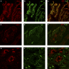
Colocalization of IgG and LRP2. (A–C) Immunofluorescence experiments performed on cryosections of normal human kidney tissue. (A) Staining for LRP2 (red) using a rabbit polyclonal antibody along the apical membrane of the tubular epithelium. (B) Indirect immunofluorescence of serum from a patient with ABBA on the normal human kidney. (C) Strong colocalization of LRP2 and serum antibodies from a patient with ABBA. (D–F) Immunofluorescence experiments performed on a renal biopsy sample from a patient with anti-LRP2 nephropathy shows granular TBM staining in a proximal tubule for LRP2 using (D) a mouse mAb and (E) IgG. (F) There is strong colocalization of LRP2 and IgG in the TBM deposits. (G–I) Immunofluorescence experiments performed on a renal biopsy sample from a patient with lupus nephritis shows staining of the tubular apical membrane for LRP2 using (G) a mouse mAb and (H) granular TBM staining for IgG. (I) There is complete lack of colocalization in a patient without anti-LRP2 nephropathy.
Figure 4.
Figure 4.
Reactivity of ABBA sera with recombinant (r) LRP2 fragments. (A) The domain structure of LRP2 is composed of four clusters of LA repeats, 17 EGF-like repeats, and eight YWTD (tyrosine-tryptophan-threonine-aspartate)-containing six-bladed β-propeller domains followed by a single transmembrane domain and a cytoplasmic tail. (B) An immunoblot under reducing conditions for the 3xFLAG tag to show the location of all four recombinant fragments. All fragments run slightly above their expected sizes of 35 kD for rLRP2(LA 1–7), 39 kD for rLRP2(LA 8–15), 50 kD for rLRP2(LA 16–25), and 32 kD for rLRP2(LA26–32). (C) ABBA serum from patient 5 is reactive with HTE and the four sets of LA repeats (nonreducing). Full-length LRP2 is close to the origin of the gel; the lower bands are proteolytic fragments that result from protease activity during preparation. (D) Sera from patients with ABBA immunoprecipitate the recombinant 3xFLAG-tagged N-terminal rLRP2(LA 1–7) fragment (lanes 1–4) under reducing conditions, whereas sera from control patients do not (lanes 5–8). The immunoprecipitated product appears distorted on the blot due to the large amount of IgG heavy chain running directly above. IP, immunoprecipitate; MW, molecular weight; WB, Western blot.

Comment in

  • Tubular disease: Anti-LRP2 nephropathy.
    Carney EF. Carney EF. Nat Rev Nephrol. 2018 Jan;14(1):3. doi: 10.1038/nrneph.2017.159. Epub 2017 Nov 13. Nat Rev Nephrol. 2018. PMID: 29129926 No abstract available.

References

    1. Chawla LS, Eggers PW, Star RA, Kimmel PL: Acute kidney injury and chronic kidney disease as interconnected syndromes. N Engl J Med 371: 58–66, 2014 - PMC - PubMed
    1. Rosales IA, Collins AB, do Carmo PA, Tolkoff-Rubin N, Smith RN, Colvin RB: Immune complex tubulointerstitial nephritis due to autoantibodies to the proximal tubule brush border. J Am Soc Nephrol 27: 380–384, 2016 - PMC - PubMed
    1. Klassen J, McCluskey RT, Milgrom F: Nonglomerular renal disease produced in rabbits by immunization with homologous kidney. Am J Pathol 63: 333–358, 1971 - PMC - PubMed
    1. Klassen J, Milgrom FM, McCluskey RT: Studies of the antigens involved in an immunologic renal tubular lesion in rabbits. Am J Pathol 88: 135–144, 1977 - PMC - PubMed
    1. Sugisaki T, Yoshida T, McCluskey RT, Andres GA, Klassen J: Autoimmune cell-mediated tubulointerstitial nephritis induced in Lewis rats by renal antigens. Clin Immunol Immunopathol 15: 33–43, 1980 - PubMed

Publication types

MeSH terms

Substances

LinkOut - more resources