. 2017 Oct 27;358(6362):535-538.
doi: 10.1126/science.aan5706.

Obstruction of pilus retraction stimulates bacterial surface sensing

Affiliations

Obstruction of pilus retraction stimulates bacterial surface sensing

Courtney K Ellison et al. Science. .

Abstract

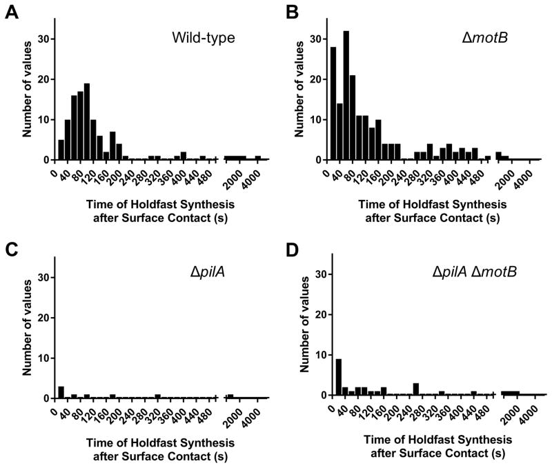
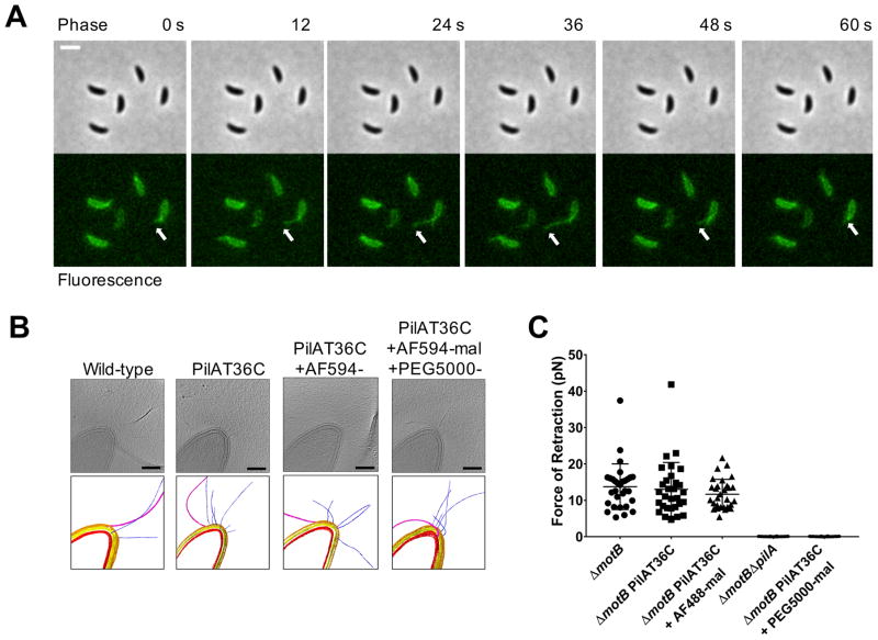
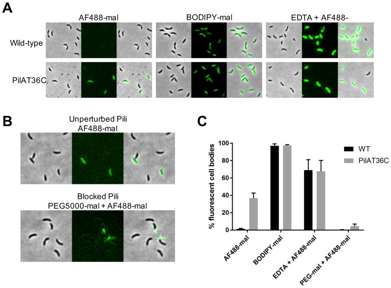
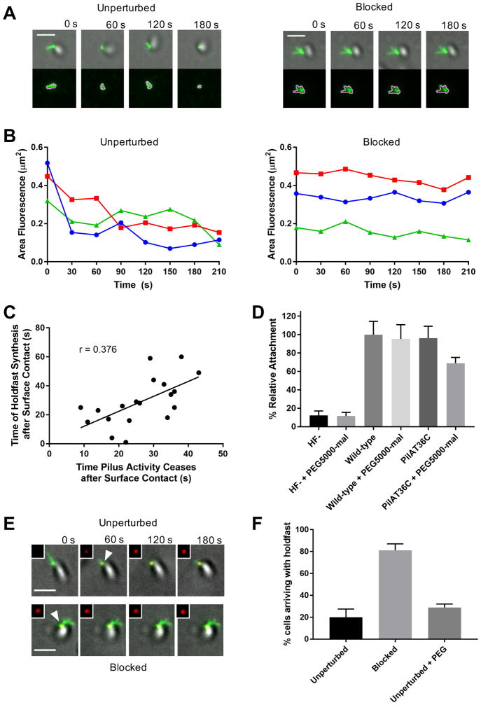
It is critical for bacteria to recognize surface contact and initiate physiological changes required for surface-associated lifestyles. Ubiquitous microbial appendages called pili are involved in sensing surfaces and facilitating downstream behaviors, but the mechanism by which pili mediate surface sensing has been unclear. We visualized Caulobacter crescentus pili undergoing dynamic cycles of extension and retraction. Within seconds of surface contact, these cycles ceased, which coincided with synthesis of the adhesive holdfast required for attachment. Physically blocking pili imposed resistance to pilus retraction, which was sufficient to stimulate holdfast synthesis without surface contact. Thus, to sense surfaces, bacteria use the resistance on retracting, surface-bound pili that occurs upon surface contact.


Figures

Figure 1
Figure 1. Tad pili are required for surface stimulation of holdfast synthesis
(A–D) Histogram plots showing the time of holdfast synthesis for single cells after surface contact for two independent replicates of wild-type (A) n = 115, ΔmotB (B) n = 182, ΔpilA (C) n = 8, and ΔpilA ΔmotB (D) n = 33. Total number of cells tracked including cells arriving with holdfast already synthesized: wild-type n = 241, ΔmotB n = 566, ΔpilA n = 93, and ΔpilAΔmotB n = 84.
Figure 2
Figure 2. Tad type IVc pili undergo dynamic cycles of extension and retraction
(A) Time-lapse of labeled, synchronized PilAT36C swarmer cells extending and retracting pili after labeling with AF488-mal dye. White arrow follows the most prominent extension and retraction event for a single cell, though all cells shown extend and retract pili. Scale bar is 2 μm. (B) Slices from tomograms and corresponding 3D segmentations of wild-type, PilAT36C, PilAT36 labeled with AF594-mal, and PilAT36C blocked with PEG5000-mal and labeled with AF594-mal. In 3D segmentation volumes, flagella are pink, pili are blue, S-layer is gold, outer membrane is yellow, and inner membrane is red. Scale bars are 200 nm. (C) Micropillars assay force measurements of retraction of tad type IVc pili in flagellar motor mutant ΔmotB strains. Flagellar motor mutants exhibiting paralyzed flagella were used to ensure all measurements obtained were dependent solely on pilus activity. Error bars show mean ± SD from 30 cells for each dataset.
Figure 3
Figure 3. C. crescentus tad pilus retraction internalizes labeled pilins into a recyclable pool of subunits
(A) Representative images of wild-type or PilAT36C cells labeled with AF488-mal, BODIPY-mal, or AF488-mal after OM permeabilization with 20 mM EDTA. (B) Representative images of PilAT36C cells labeled with AF488-mal ± PEG5000-mal. (C) Quantification of fluorescent cell bodies in populations of cells from images shown in (A and B). A minimum of 398 cells from each of 3 independent biological replicates was quantified. Error bars show mean ± SD.
Figure 4
Figure 4. Resistance to C. crescentus tad pilus retraction triggers surface stimulation of holdfast synthesis
(A) Representative TIRF images of unperturbed cell labeled with AF488-mal exhibiting dynamic pilus activity and blocked cell labeled with both AF488-mal and PEG5000-mal exhibiting no dynamic pilus activity with overlays of gray cell body and green fluorescent pili. The bottom image panel shows green fluorescent pili surrounded by pink MicrobeJ overlay used to measure changes in fluorescence area of pili (μm2) over time shown in graphs (B). (B) Graphs showing changes in fluorescence area occupied by pili over time by cells shown in (A). (C) Plot showing correlation between the time of holdfast synthesis after surface contact and the time of cessation of dynamic pilus activity after surface contact for 19 cells. (D) Relative attachment assay showing binding efficiency of holdfast minus (HF-) and PilAT36C strains compared to wild-type after 30 min binding ± PEG5000-mal. Data are representative of binding from 3 independent cultures normalized to wild-type binding levels. Error bars show mean ± SD. PilAT36C + PEG-mal is significantly different from all other treatments based on unpaired, two-tailed T-test (P < 0.03). (E) Representative TIRF microscopy images of cells unperturbed or blocked for pilus retraction upon surface contact in the presence of AF594-WGA where time = 0 s is time of surface contact. The cell body is gray, labeled pili are green, and HF are red, and shown in upper right inset of each image. White arrowheads represent first appearance of holdfast for cell depicted. Scale bars are 2 μm. (F) Quantification of cells after labeling with AF488-mal (unperturbed); AF488-mal + PEG5000-mal (blocked); or AF488-mal + PEG5000 (unperturbed + PEG). A minimum of 30 cells from each of 3 independent biological replicates was quantified. Error bars show mean ± SD. Scale bars are 2 μm.

Comment in

  • The bacterium has landed.
    Hughes KT, Berg HC. Hughes KT, et al. Science. 2017 Oct 27;358(6362):446-447. doi: 10.1126/science.aaq0143. Science. 2017. PMID: 29074753 Free PMC article. No abstract available.

References

    1. Hoffman MD, et al. Timescales and Frequencies of Reversible and Irreversible Adhesion Events of Single Bacterial Cells. Anal Chem. 2015 doi: 10.1021/acs.analchem.5b02087. - DOI - PMC - PubMed
    1. Li G, et al. Surface contact stimulates the just-in-time deployment of bacterial adhesins. Mol Microbiol. 2012;83:41–51. - PMC - PubMed
    1. Utada AS, et al. Vibrio cholerae use pili and flagella synergistically to effect motility switching and conditional surface attachment. Nat Commun. 2014;5:4913. - PMC - PubMed
    1. Siryaporn A, Kuchma SL, O’Toole GA, Gitai Z. Surface attachment induces Pseudomonas aeruginosa virulence. Proc Natl Acad Sci U S A. 2014;111:16860–5. - PMC - PubMed
    1. Persat A, Inclan YF, Engel JN, Stone HA, Gitai Z. Type IV pili mechanochemically regulate virulence factors in Pseudomonas aeruginosa. Proc Natl Acad Sci. 2015;112:7563–7568. - PMC - PubMed

Publication types

MeSH terms

Substances