. 2017 Oct 26;7(1):14133.
doi: 10.1038/s41598-017-14207-0.

Differentiation of Oligodendrocyte Precursor Cells from Sox10-Venus Mice to Oligodendrocytes and Astrocytes

Affiliations

Differentiation of Oligodendrocyte Precursor Cells from Sox10-Venus Mice to Oligodendrocytes and Astrocytes

Nobuharu Suzuki et al. Sci Rep. .

Abstract

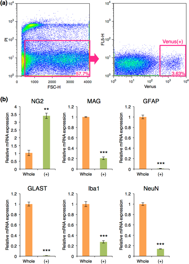
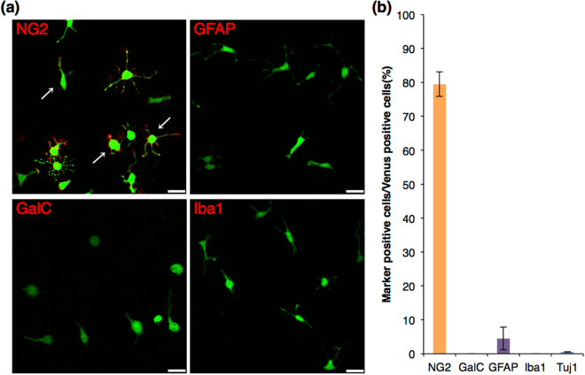
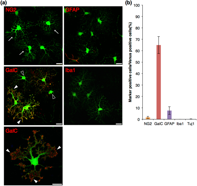
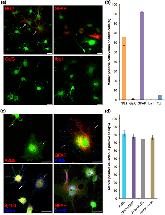
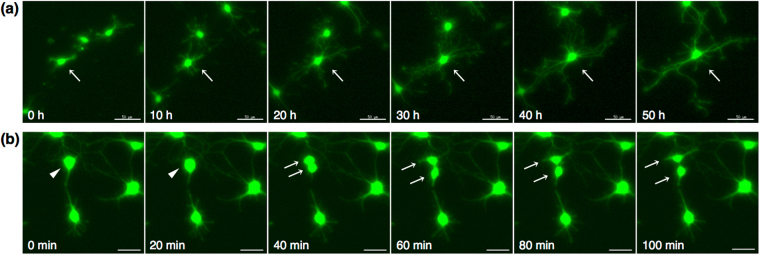
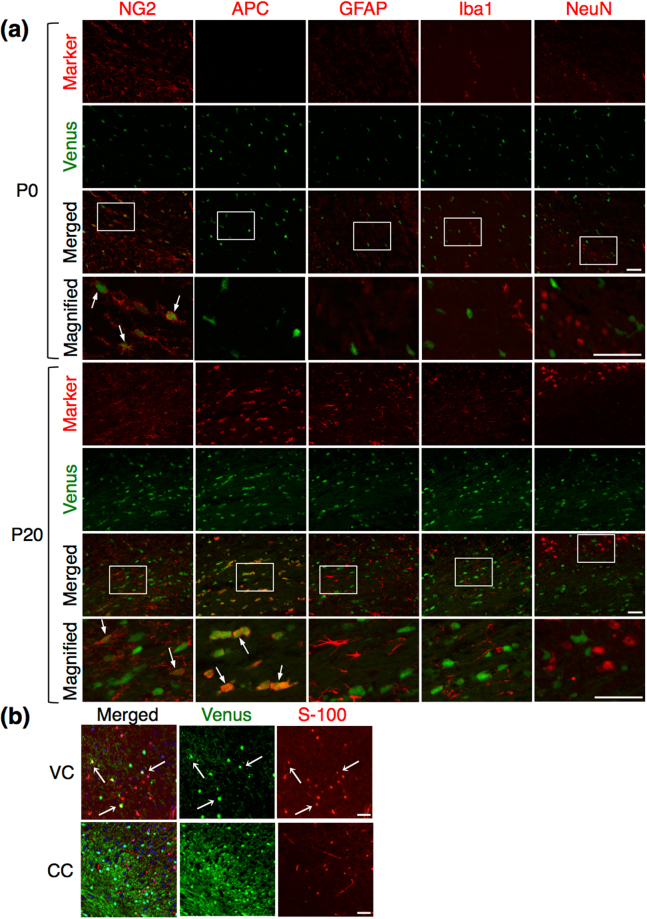
Oligodendrocytes are well known as myelin-forming cells in the central nervous system (CNS). However, detailed mechanisms of oligodendrocyte differentiation and myelination are poorly understood, particularly due to the difficulty of the purification of murine oligodendrocyte precursor cells (OPCs). We have recently established a transgenic mouse line that expresses a fluorescent protein Venus under the promoter of Sox10, whose expression is restricted to OPCs and oligodendrocytes in the CNS. Here, we have characterized Venus-positive cells from the Sox10-Venus mouse brain for analyzing oligodendrocyte differentiation. We first purified Venus-positive cells from the postnatal day 0-2 brain by flow cytometry. Most of the Venus-positive cells expressed NG2, an OPC marker. After induction of differentiation, an increased population of galactocerebroside-positive oligodendrocytes and decrease of OPCs were observed in the Venus-positive culture. Furthermore, a time-lapse analysis showed that Venus-positive oligodendrocytes dynamically changed their morphology with highly branched cell processes during differentiation. In addition, we found that Venus-positive OPCs were able to differentiate to type II astrocytes. In vivo, OPCs and oligodendrocytes express Venus, and some of astrocytes were positive for Venus in the ventral cortex. Taken together, the Sox10-Venus mouse system is useful for analyzing differentiation and multipotency of murine OPCs.


Conflict of interest statement

The authors declare that they have no competing interests.

Figures

Figure 1
Figure 1
Cell sorting of Venus (+) cells using flow cytometry. (a) Flow cytometry analysis of Venus (+) cells. Cells positive for propidium iodide were removed as dead cells (pink box in the left panel), and cells with the strong intensity of Venus were collected as Venus (+) cells (pink box in the right panel). The numbers in pink indicates the fraction of each cell population in the flow cytometry. (b) Quantitative RT-PCR of glial and neural markers in whole brain cells before cell sorting and Venus (+) cells. The mRNA expression of each gene was normalized using β-actin mRNA expression. The normalized expression of each gene in whole brain cells was set as 1.0. Whole: whole brain cells before cell sorting, orange bar; (+): Venus (+) cells, green bar; Error bars, s.e.m. (**p < 0.01, ***p < 0.001, t test).
Figure 2
Figure 2
Immunocytochemistry of glial and neuronal markers in Venus (+) cells before induction of differentiation. (a) Confocal images of the immunocytochemistry before induction of differentiation. Venus fluorescence is shown in green, and each glial marker is shown in red. Scale bar, 25 µm. Venus (+) cells expressed NG2, showing typical cell morphology of OPCs in culture (arrows). (b) Cell count analysis showing cell population of Venus (+) cells expressing each marker. Error bars, s.e.m.
Figure 3
Figure 3
Immunocytochemistry of glial and neuronal markers in Venus (+) cells after oligodendrocyte differentiation. (a) Confocal images of the immunocytochemistry after oligodendrocyte differentiation. Venus fluorescence is shown in green, and each glial marker is shown in red. Venus (+) OPCs formed highly branched cell processes after 3 days of culture in Serum-free differentiation medium (arrows). These cells expressed GalC, showing sheet-like structures in their cellular processes (closed arrowheads). GalC-negative cells, whose morphology was likely differentiating OPCs to oligodendrocytes, were observed (opened arrowheads). Scale bar, 25 µm. (b) Cell count analysis showing cell population of Venus (+) cells expressing each marker. Error bars, s.e.m.
Figure 4
Figure 4
Immunocytochemistry of glial and neuronal markers in Venus (+) cells after astrocytic differentiation. (a,c) Confocal images of the immunocytochemistry after astrocytic differentiation. Venus fluorescence is shown in green, and each glial marker is shown in red or blue. Venus (+) OPCs developed into flat shaped cells with short primary cell processes (A: arrows). Most of Venus (+) cells expressed A2B5 with GFAP or S-100, or GFAP with S-100. Arrow: punctated staining of A2B5 on Venus (+) cell; Scale bar, 25 µm. (b,d) Cell count analysis showing cell population of Venus (+) cells expressing each marker. Error bars, s.e.m.
Figure 5
Figure 5
Time-lapse analysis of Venus (+) cells. (a) Differentiation of Venus (+) OPCs. In vitro time-lapse images were captured to follow the process formation of Venus (+) oligodendrocytes after induction of differentiation. Images every 10 hours are representatively indicated. Arrow: differentiating OPC with branched process formation; Scale bar, 50 µm. (b) Cell division of Venus (+) OPCs. Representative cell division images are shown every 20 minutes. Arrowhead: OPC before cell division; Arrows: OPCs after cell division; Scale bar, 30 µm.
Figure 6
Figure 6
Immunohistochemistry of glial and neuronal markers in Venus (+) cells in the brain tissues. (a) Immunohistochemistry in the corpus callosum of the P0 and P20 posterior forebrain, showing the distribution of Venus positive cells (green) with each marker (red). Arrows: dual-positive cells of Venus with a marker; Scale bar: 50 µm. (b) Immunohistochemistry of S-100 in the ventral cortex (VC) and the corpus callosum (CC) at 12 months. Dual-positive cells of Venus with S-100 were observed in the ventral cortex (arrows); Scale bar: 50 µm.

References

    1. Boulanger JJ, Messier C. From precursors to myelinating oligodendrocytes: contribution of intrinsic and extrinsic factors to white matter plasticity in the adult brain. Neuroscience. 2014;269:343–366. doi: 10.1016/j.neuroscience.2014.03.063. - DOI - PubMed
    1. Nishiyama A, Komitova M, Suzuki R, Zhu X. Polydendrocytes (NG2 cells): multifunctional cells with lineage plasticity. Nat. Rev. Neurosci. 2009;10:9–22. doi: 10.1038/nrn2495. - DOI - PubMed
    1. Nishiyama A, Suzuki R, Zhu X. NG2 cells (polydendrocytes) in brain physiology and repair. Front Neurosci. 2014;8:133. doi: 10.3389/fnins.2014.00133. - DOI - PMC - PubMed
    1. Behar TN. Analysis of fractal dimension of O2A glial cells differentiating in vitro. Methods. 2001;24:331–339. doi: 10.1006/meth.2001.1203. - DOI - PubMed
    1. Chen Y, et al. Isolation and culture of rat and mouse oligodendrocyte precursor cells. Nat. Protoc. 2007;2:1044–1051. doi: 10.1038/nprot.2007.149. - DOI - PubMed

Publication types

LinkOut - more resources