. 2017 Oct 26;7(1):13476.
doi: 10.1038/s41598-017-14003-w.

Motor recovery after activity-based training with spinal cord epidural stimulation in a chronic motor complete paraplegic

Affiliations

Motor recovery after activity-based training with spinal cord epidural stimulation in a chronic motor complete paraplegic

Enrico Rejc et al. Sci Rep. .

Abstract

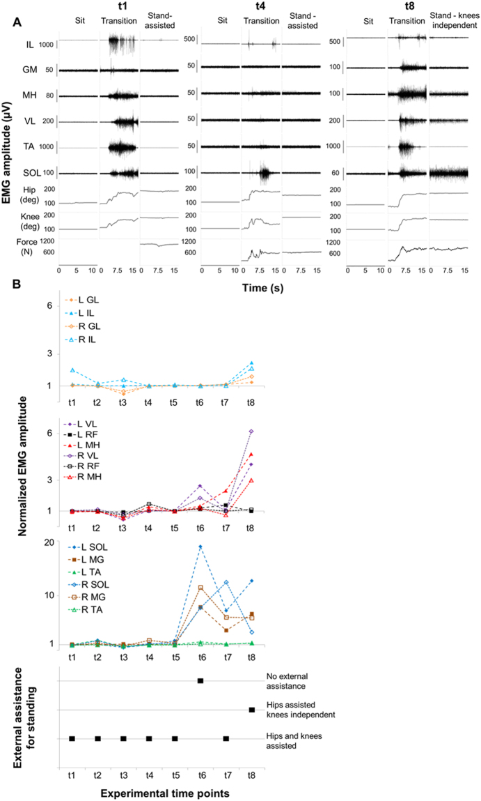
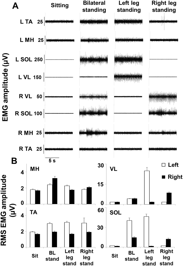
The prognosis for recovery of motor function in motor complete spinal cord injured (SCI) individuals is poor. Our research team has demonstrated that lumbosacral spinal cord epidural stimulation (scES) and activity-based training can progressively promote the recovery of volitional leg movements and standing in individuals with chronic clinically complete SCI. However, scES was required to perform these motor tasks. Herein, we show the progressive recovery of voluntary leg movement and standing without scES in an individual with chronic, motor complete SCI throughout 3.7 years of activity-based interventions utilizing scES configurations customized for the different motor tasks that were specifically trained (standing, stepping, volitional leg movement). In particular, this report details the ongoing neural adaptations that allowed a functional progression from no volitional muscle activation to a refined, task-specific activation pattern and movement generation during volitional attempts without scES. Similarly, we observed the re-emergence of muscle activation patterns sufficient for standing with independent knee and hip extension. These findings highlight the recovery potential of the human nervous system after chronic clinically motor complete SCI.


Conflict of interest statement

The authors declare that they have no competing interests.

Figures

Figure 1
Figure 1
Experimental protocol timeline. Panel (A) Standard of care and outpatient rehabilitation (locomotor training, LT) within the initial 21 months since injury. Panel (B) Experimental sessions (t a to t8) and activity-based interventions performed prior to and after scES implant (see text for details).
Figure 2
Figure 2
Quantitative joint probability density distributions analysis. The scatterplot obtained by the joint probability density distributions analysis (Hutchison et al.), which describes the amplitude and temporal interrelationships of the EMG signals from two muscles (Ma and Mb), was divided into four areas (A,B,C and D) that were determined by setting a threshold equal to 10% of the of the EMG amplitude full scale values.
Figure 3
Figure 3
Volitional attempts to perform hip flexion. Panel (A) Electromyography (EMG) activity and hip joint angle recorded during representative attempts to volitionally perform right hip flexion in the supine position without scES after 80 sessions of locomotor training without scES (t1), after 9.5 months (t3) and after 44 months (t8) of activity-based training with scEs (see Fig. 1 for details). Panel (B) Probability density distribution of EMG amplitudes between the iliopsoas (IL, hip flexor) and the medial hamstrings (MH, hip extensors) calculated during the volitional attempts (data comprised between the two vertical grey dotted lines in Panel A) performed at the experimental time points t1, t3 and t8. Panel (C) EMG amplitude recorded during the volitional attempts, normalized by background (resting) EMG amplitude, and the resulting amount of hip flexion. Kinematics was not recorded at experimental time point t a. Panel (D) Quantitative probability density distribution of EMG amplitudes between iliopsoas and medial hamstrings calculated during the volitional attempts. Black and green indicate the amount of co-contraction at low or high level of activation, respectively (area A and D showed in Fig. 2). Blue and red indicate the isolate activation of iliopsoas or medial hamstrings, respectively (area C and B showed in Fig. 2). EMG was recorded from the following muscles of the right lower limb: IL, iliopsoas; GL, gluteus maximus; MH, medial hamstring; VL, vastus lateralis; TA, tibialis anterior; SOL, soleus. At the experimental time point t a, rectus femoris (RF) was monitored instead of IL as representative hip flexor. EMG activity from intercostal (IC) muscle was recorded to monitor the volitional effort onset.
Figure 4
Figure 4
Volitional attempts to perform knee extension. Panel (A) Electromyography (EMG) activity recorded during representative attempts to volitionally perform right knee extension in the supine position without scES after 80 sessions of locomotor training without scES (t b), after 9.5 months (t3) and after 44 months (t8) of activity-based training with scEs (see Fig. 1 for details). Panel (B) Probability density distribution of EMG amplitudes between the vastus lateralis (VL, knee extensor) and the medial hamstrings (MH, knee flexors) calculated during the volitional attempts (data comprised between the two vertical grey dotted lines in Panel A) performed at the experimental time points t b, t3 and t8. Panel (C) EMG amplitude recorded during the volitional attempts, normalized by background (resting) EMG amplitude, and the resulting knee joint movement. Panel (D) Quantitative probability density distribution of EMG amplitudes between the vastus lateralis and the medial hamstrings calculated during the volitional attempts. Black and green indicate the amount of co-contraction at low or high level of activation, respectively (area A and D showed in Fig. 2). Blue and red indicate the isolate activation of vastus lateralis or medial hamstrings, respectively (area C and B showed in Fig. 2). EMG was recorded from the following muscles of the right lower limb: RF, rectus femoris; VL, vastus lateralis; MH, medial hamstring; TA, tibialis anterior; SOL, soleus. EMG activity from sternocleidomastoid (SCM) muscle was recorded to monitor the volitional effort onset.
Figure 5
Figure 5
Time course of EMG amplitude and external assistance during standing. Panel (A) Electromyography (EMG) activity, hip and knee joint angle, and ground reaction forces recorded during sitting, sit-to-stand transition and overground full weight-bearing standing without epidural stimulation prior to any training (t1), after 15.5 months (t4) and after 44 months (t8) of activity-based training with scEs (see Fig. 1 for details). Panel (B) Time course of EMG amplitude recorded during standing, normalized by background (resting) EMG amplitude, and amount of external assistance needed for standing without epidural stimulation. EMG was recorded from the following muscles of the left (L) and right (R) lower limb: IL, iliopsoas; GL, gluteus maximus; MH, medial hamstring; VL, vastus lateralis; RF: rectus femoris; TA, tibialis anterior; SOL, soleus; MG: medial gastrocnemius.
Figure 6
Figure 6
EMG activity during bilateral and unilateral independent standing. Panel (A) Electromyography (EMG) activity recorded during sitting, bilateral and unilateral full weight-bearing independent standing without epidural stimulation after 29.5 months of activity-based training with scEs (t6, see Fig. 1 for details). Panel (B) EMG amplitude calculated as the root mean square (RMS) over 10 seconds of steady sitting, bilateral and unilateral independent standing. EMG was recorded from the left (L) and right (R) medial hamstring (MH), vastus lateralis (VL), tibialis anterior (TA) and soleus (SOL).

References

    1. Varma AK, et al. Spinal cord injury: a review of current therapy, future treatments, and basic science frontiers. Neurochem. Res. 2013;38(5):895. doi: 10.1007/s11064-013-0991-6. - DOI - PMC - PubMed
    1. Waters RL, et al. Recovery following complete paraplegia. Arch. Phys. Med. Rehabil. 1992;73(9):784. - PubMed
    1. Colombo G, Wirz M, Dietz V. Effect of locomotor training related to clinical and electrophysiological examinations in spinal cord injured humans. Ann. N. Y. Acad. Sci. 1998;860:536. doi: 10.1111/j.1749-6632.1998.tb09097.x. - DOI - PubMed
    1. Dietz V, Colombo G, Jensen L. Locomotor activity in spinal man. Lancet. 1994;344(8932):1260. doi: 10.1016/S0140-6736(94)90751-X. - DOI - PubMed
    1. Dietz V, et al. Locomotor capacity of spinal cord in paraplegic patients. Ann. Neurol. 1995;37(5):574. doi: 10.1002/ana.410370506. - DOI - PubMed

Publication types