Review
. 2017:2017:5035371.
doi: 10.1155/2017/5035371. Epub 2017 Sep 17.

Desired Turbulence? Gut-Lung Axis, Immunity, and Lung Cancer

Affiliations
Review

Desired Turbulence? Gut-Lung Axis, Immunity, and Lung Cancer

Rea Bingula et al. J Oncol. 2017.

Abstract

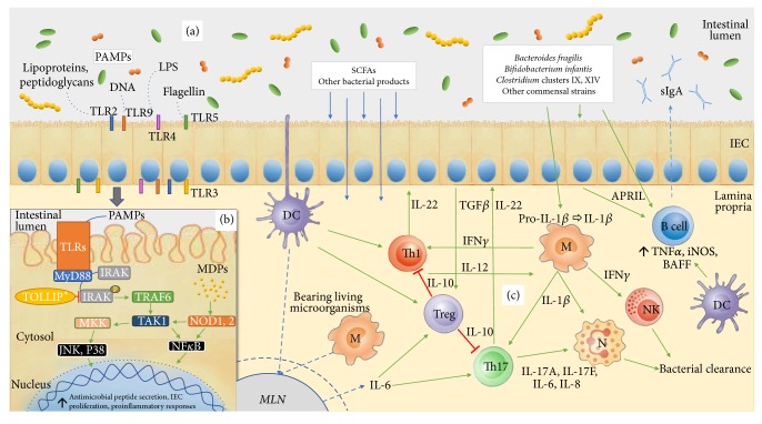
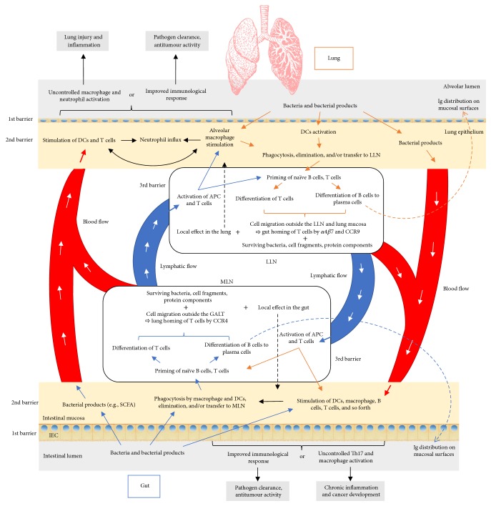
The microbiota includes different microorganisms consisting of bacteria, fungi, viruses, and protozoa distributed over many human body surfaces including the skin, vagina, gut, and airways, with the highest density found in the intestine. The gut microbiota strongly influences our metabolic, endocrine, and immune systems, as well as both the peripheral and central nervous systems. Recently, a dialogue between the gut and lung microbiota has been discovered, suggesting that changes in one compartment could impact the other compartment, whether in relation to microbial composition or function. Further, this bidirectional axis is evidenced in an, either beneficial or malignant, altered immune response in one compartment following changes in the other compartment. Stimulation of the immune system arises from the microbial cells themselves, but also from their metabolites. It can be either direct or mediated by stimulated immune cells in one site impacting the other site. Additionally, this interaction may lead to immunological boost, assisting the innate immune system in its antitumour response. Thus, this review offers an insight into the composition of these sites, the gut and the lung, their role in shaping the immune system, and, finally, their role in the response to lung cancer.


Figures

Figure 1
Figure 1
Interaction of the microbiota and intestinal mucosa. (a) Microorganisms in the intestine provide pathogen-associated molecular patterns (PAMPs) that serve as ligands for different Toll-like receptors (TLRs) on the luminal or basolateral surface of the intestinal epithelial cells (IECs). (b) TLRs stimulation activates a signalling cascade resulting in transcription factor activation and gene transcription, enhancing the cell barrier and further stimulating the immunological cells in the lamina propria. This cascade can be inhibited by toll-interacting protein (TOLLIP). Inhibition seen only for TLR2 and TLR4 [23]. (c) Commensal bacteria and their derivatives (e.g., short-chain fatty acids (SCFAs)) directly stimulate IECs (b) or can be phagocytosed by DCs and macrophages in lamina propria and carried to mesenteric lymph nodes (MLN) where they prime naïve B and T cells to mature and differentiate. B cells become plasma cells and produce IgA that is secreted into the intestinal lumen (sIgA). T cells profile into Th17 and Th1, with proinflammatory tendency, activating additional effector cells as neutrophils, resulting in bacterial clearance. There is also differentiation to Treg cells having anti-inflammatory properties and controlling inflammation. APRIL: a proliferation-inducing ligand; BAFF: B cell-activating factor of the tumour necrosis family; DC: dendritic cell; IEC: intestinal epithelial cells; IFN: interferon; IL: interleukin; iNOS: inducible nitric oxide synthase; IRAK: interleukin receptor-associated kinase; JNK: c-Jun N-terminal kinases; LLN: lung lymph node; LPS: lipopolysaccharide; M: macrophage; MDPs: microbiota derived particles; MKK: mitogen-activated protein kinase kinase; MLN: mesenteric lymph node; MyD88: myeloid differentiation primary response gene 88; N: neutrophil; NF-κB: nuclear factor kappa-light-chain-enhancer of activated B cells; NK: natural killer cell; NOD: nucleotide-binding receptor; PAMPs: pathogen-associated molecular patterns; SCFAs: short-chain fatty acids; TAK-1: transforming growth factor beta-activated kinase; TGFβ: transforming growth factor beta; Th: T-helper cell; TLR: toll-like receptor; TNFα: tumour necrosis factor alfa; TOLLIP: toll-interacting protein; TRAF6: TNF receptor-associated factor 6; Treg: regulatory T cell.
Figure 2
Figure 2
Proposed pathways of the gut-lung interaction. Microbiota and its products that enter intestinal mucosa (blue arrows) are phagocytosed and transferred to mesenteric lymph nodes (MLN) by antigen presenting cells (APC), where they stimulate priming of the T and B cells. Once activated, with the expression of proper homing receptors, these cells can migrate back to the original site (intestinal mucosa) (black dashed arrow) or to distal locations such as the lung epithelium and lung nodes through lymphatic and blood circulation. There, they can directly act on their target or continue to stimulate other immune cells. On the other hand, bacterial products from the intestinal mucosa or surviving bacteria can also reach the lung by blood or lymphatics to stimulate the immune system in the same way as they would have done in the intestinal tract. Depending on the tissue prestimulation, type of stimulus, and local and general immunological status, the result can be positive, as effective bacterial clearance or antitumour activity, or overinflammatory response, promoting further tissue damage, pathogen colonization, and tumour progression. The same schema is proposed in the other sense, beginning with the lung mucosa and finishing with distal effect on the gut. Although not known for the moment, there is also a possibility that bacterial products of the lung microbiota can exert their effect in the intestinal mucosa, being delivered in the same way as explained above. APC: antigen presenting cell; DC: dendritic cell; GALT: gut-associated lymphatic tissue; IEC: intestinal epithelial cell; LLN: lung lymph node; MLN: mesenteric lymph node; SCFA: short-chain fatty acid. Colour legend (borders/arrows): blue: influence of gut on lung; orange: influence of lung on gut; black: mutual influence.

References

    1. Marsland B. J., Trompette A., Gollwitzer E. S. The gut-lung axis in respiratory disease. Annals of the American Thoracic Society. 2015;12(Suppl 2):S150–S156. doi: 10.1513/AnnalsATS.201503-133AW. - DOI - PubMed
    1. Brownawell A. M., Caers W., Gibson G. R., et al. Prebiotics and the health benefits of fiber: Current regulatory status, future research, and goals. Journal of Nutrition. 2012;142(5):962–974. doi: 10.3945/jn.112.158147. - DOI - PubMed
    1. Belkaid Y., Hand T. W. Role of the microbiota in immunity and inflammation. Cell. 2014;157(1):121–141. doi: 10.1016/j.cell.2014.03.011. - DOI - PMC - PubMed
    1. Carding S., Verbeke K., Vipond D. T., Corfe B. M., Owen L. J. Dysbiosis of the gut microbiota in disease. Microbial Ecology in Health & Disease. 2015;26(1) doi: 10.3402/mehd.v26.26191. - DOI - PMC - PubMed
    1. Forsythe P., Kunze W., Bienenstock J. Moody microbes or fecal phrenology: What do we know about the microbiota-gut-brain axis? BMC Medicine. 2016;14(1, article no. 58) doi: 10.1186/s12916-016-0604-8. - DOI - PMC - PubMed

LinkOut - more resources