. 2017 Oct 19:14:48.
doi: 10.1186/s12983-017-0234-9. eCollection 2017.

The flipflop orphan genes are required for limb bud eversion in the Tribolium embryo

Affiliations

The flipflop orphan genes are required for limb bud eversion in the Tribolium embryo

Susanne Thümecke et al. Front Zool. .

Abstract

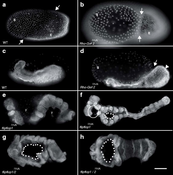
Background: Unlike Drosophila but similar to other arthropod and vertebrate embryos, the flour beetle Tribolium castaneum develops everted limb buds during embryogenesis. However, the molecular processes directing the evagination of epithelia are only poorly understood.

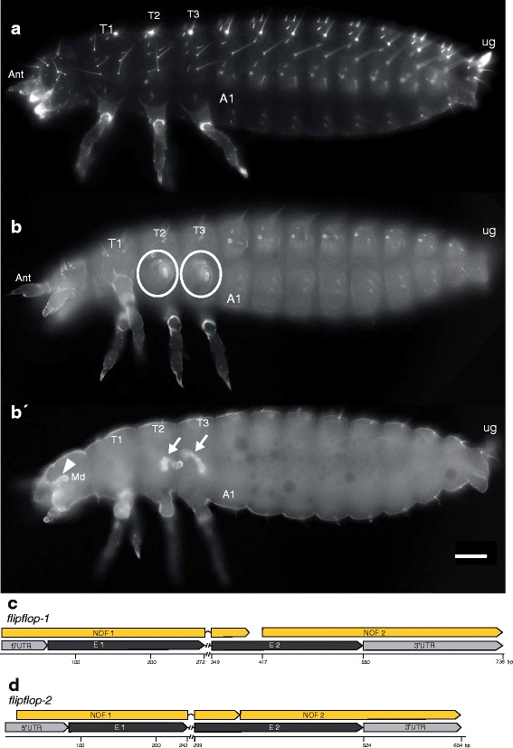
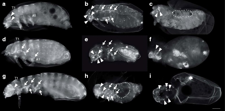
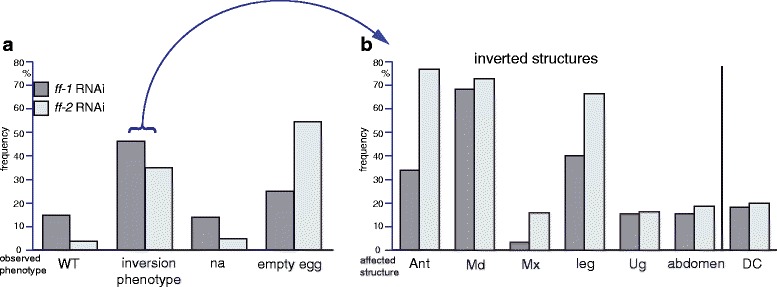
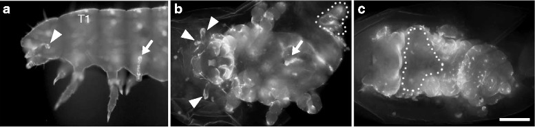
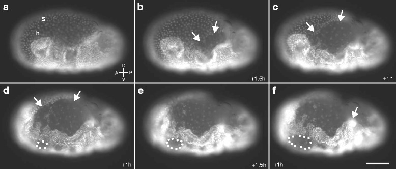
Results: Here we show that the newly discovered genes Tc-flipflop1 and Tc-flipflop2 are involved in regulating the directional budding of appendages. RNAi-knockdown of Tc-flipflop results in a variety of phenotypic traits. Most prominently, embryonic limb buds frequently grow inwards rather than out, leading to the development of inverted appendages inside the larval body. Moreover, affected embryos display dorsal closure defects. The Tc-flipflop genes are evolutionarily non-conserved, and their molecular function is not evident. We further found that Tc-RhoGEF2, a highly-conserved gene known to be involved in actomyosin-dependent cell movement and cell shape changes, shows a Tc-flipflop-like RNAi-phenotype.

Conclusions: The similarity of the inverted appendage phenotype in both the flipflop- and the RhoGEF2 RNAi gene knockdown led us to conclude that the Tc-flipflop orphan genes act in a Rho-dependent pathway that is essential for the early morphogenesis of polarised epithelial movements. Our work describes one of the few examples of an orphan gene playing a crucial role in an important developmental process.

Keywords: Appendage formation; Epithelial morphogenesis; Evagination; Orphan flipflop gene; PCP; RhoGEF2; Tissue folding; Tribolium castaneum.


Conflict of interest statement

Ethics approval and consent to participate

Not applicable

Consent for publication

Not applicable

Competing interests

The authors declare that they have no competing interests.

Publisher’s Note

Springer Nature remains neutral with regard to jurisdictional claims in published maps and institutional affiliations.

Figures

Fig. 1
Fig. 1
Larval Tc-flipflop RNAi phenotype. (a) Wildtype cuticle. All appendages develop as everted structures. Each thoracic segment bears one pair of visible legs. (b-b′) Tc-ff RNAi phenotype. (b) Surface view, () optical section. (b) In the thoracic segments 2 and 3, one leg is everted normally. Of the other legs, only the coxa is visible as an outer structure (circle) while the remaining leg is internalised (b′, arrows). (c, d) Gene organisation of Tc-ff1 (c) and Tc-ff2 (d) genes including NOF (non-overlapping-fragment) positions used for RNAi experiments. A1 abdominal segment 1; Ant antenna; Md mandible; T1–3 thoracic segment 1–3; ug urogomphi; scale bar (a-b′) 100 μm; all panels in all pictures: anterior to the left
Fig. 2
Fig. 2
Detailed view of the larval Tc-flipflop RNAi phenotype. a, b Magnified view of a larval inverted antenna a and leg b. Internalised parts are fully differentiated; the distalmost structures (antennal flagellum) (arrowhead in a) and the pretarsal claw (arrow in b) develop normally and display the inside-out event. Scalebar 20 μm
Fig. 3
Fig. 3
The “flipflop syndrome”. a-i Larval RNAi phenotypes of Tc-ff1 a-c, Tc-ff2 d-f and Tc-ff1/ff2 g-i parental double-knockdown. a-i The enigmatic phenotype of inverted head appendages (arrowhead) and legs (arrows) in different combinations. In addition, inverted abdominal segments (e, circle) and dorsal openings (c, h, dotted line) are detected. f, i In stronger cuticle phenotypes cuticle remnants also show inverted appendages. A1 abdominal segment 1; T1 thoracic segment 1; scale bar 100 μm; all panels in all pictures: anterior to the left
Fig. 4
Fig. 4
Quantitative analysis of Tc-ff1 and Tc-ff2 phenotypes a Classification of cuticle phenotypes into wildtype-like (WT), analysable cuticles with inversions, not analysable cuticles (na) and “empty eggs” without visible cuticle within the vitelline membrane. b Detailed description of the “Analysable cuticles”-class (Arrow from A to B) subdivided into inversion events of antenna (Ant), mandible (Md), maxilla (Mx), leg, urogomphi (Ug) and abdominal segments as well as cuticles with a dorsal opening (DC). Ant antenna; DC dorsal closure; Md mandible; Mx maxilla; na not analysable; Ug urogomphi; WT wildtype
Fig. 5
Fig. 5
Marker gene expression and apoptosis analysis in Tc-ff RNAi embryos. Ventral focal plane: a, b, b´´, b, d, e-f´. dorsal focal plane: b´, d´. a-b´´: Tc-drumstick; c-: Tc-Distalless; e-: apoptosis-marker anti-Dcp1. (a, b-b″) Tc-drumstick (Tc-drm; TC006347) marker gene expression in embryonic wildtype (WT) and Tc-ff RNAi embryos marks the segmental base of appendages. (b, b′) Inverted mandible clearly displays wildtype Tc-drm expression at the dorsal focal plane (b′). (c, d, d’) Tc-Distalless (Tc-Dll) marker gene expression in embryonic wildtype (c) and Tc-ff RNAi embryos (d, d’) marks the distal portion of each appendage except for the mandible. (d) Some leg anlagen are not visible in the ventral focal plane (circles) but are visible as inverted limb buds in the dorsal focal plane (arrows) (d’). (e-f) Apoptosis marker anti-Dcp1 in wildtype (e) and Tc-ff RNAi embryos (f-f′). (d) Inverted appendage buds do not show elevated cell death at the stage of bud formation (circles). (f′) Nuclear staining (DAPI) visualises embryonic morphology of inverted appendages after Tc-ff knockdown. A1 abdominal segment 1; Lb labium; Md mandible; Mx maxilla; T1 thoracic segment 1; scale bar 100 μm; all panels in all pictures: anterior to the left
Fig. 6
Fig. 6
Larval Tc-RhoGEF2 RNAi phenotype. a Magnified view of the KT221 mutant phenotype displaying an inverted leg (arrow) and head appendage (arrowhead). b, c RhoGEF2 RNAi cuticles display more severe defects: in addition to inverted appendages of the head (arrowheads) and thorax (arrows) RhoGEF2 knockdown also results in segmentation defects, a malformed posterior abdomen (b dotted outline) and dorsal closure defects (c, outlined with dots). Extended focus in all pictures combines several optical sections. a lateral view; b ventral view; c dorsal view); b, c scale bar 100 μm
Fig. 7
Fig. 7
flipflop- and RhoGEF2-RNAi knockdown interferes with early embryonic morphogenesis. a Wildtype blastoderm stage displays distinction between anterior extraembryonic tissue-anlagen (serosa S) and the posterior positioned embryonic cells (E); arrows mark the border. b Fixed RNAi embryos reveal tissue constrictions in blastoderm stages during embryonic anlagen formation (arrows). c Wildtype embryo prior to axis elongation. d Young RhoGEF2 RNAi embryo with thickened posterior growth zone (arrowhead). The affected embryo is excluded from the serosa (arrow). e, h Extended germband displays an unusual S-like orientation within the yolk (y). e, inverted head appendages (f, circle) appear missing when viewed ventrally. f, g, h Median holes in ff1- and ff1/RhoGEF2 RNAi embryos (dotted line). a, c, d, e Lateral view; f-g ventral view; scale bar 100 μm; all panels in all pictures: anterior to the left; Nuclear DAPI staining
Fig. 8
Fig. 8
Live imaging stills of an Tc-ff/RhoGEF2 double-RNAi embryo. a-f Developmental dynamics in a timeframe of 6 h, starting a few hours after egg lay. a Germ anlage covered by the intact serosa (S). b-f Rupture and successive retraction of extraembryonic membranes (arrows). d-e Holes form in the median regions of the embryo (dotted outline) and ingression of embryonic tissue into the yolk (arrow in f). a-f Lateral view; anterior to the left, ventral down; scale bar 100 μm

References

    1. Alva V, Nam SZ, Söding J, Lupas AN. The MPI bioinformatics toolkit as an integrative platform for advanced protein sequence and structure analysis. Nucleic Acids Res. 2016;44:W410–W415. doi: 10.1093/nar/gkw348. - DOI - PMC - PubMed
    1. Angelini DR, Kaufman TC. Insect appendages and comparative ontogenetics. Dev Biol. 2005;286:57–77. doi: 10.1016/j.ydbio.2005.07.006. - DOI - PubMed
    1. Barrett K, Leptin M, Settleman J. The Rho GTPase and a putative RhoGEF mediate a signaling pathway for the cell shape changes in Drosophila gastrulation. Cell. 1997;91:905–15. - PubMed
    1. Beermann A, Jay DG, Beeman RW, Hülskamp M, Tautz D, Jürgens G. The Short antennae gene of Tribolium is required for limb development and encodes the orthologue of the Drosophila Distal-less protein. Development. 2001;128:287–97. - PubMed
    1. Brown, SJ, Shippy, TD, Miller, S, Bolognesi, R, Beeman, RW, Lorenzen, MD, Bucher, G, Wimmer, EA & Klingler, M. The Red Flour Beetle, Tribolium castaneum (Coleoptera): A Model for Studies of Development and Pest Biology. Cold Spring Harb Protoc 2009, doi:https://doi.org/10.1101/pdb.emo126. - DOI - PubMed