. 2017:2:17024.
doi: 10.1038/natrevmats.2017.24. Epub 2017 May 9.

Rethinking cancer nanotheranostics

Affiliations

Rethinking cancer nanotheranostics

Hongmin Chen et al. Nat Rev Mater. 2017.

Abstract

Advances in nanoparticle synthesis and engineering have produced nanoscale agents affording both therapeutic and diagnostic functions that are often referred to by the portmanteau 'nanotheranostics'. The field is associated with many applications in the clinic, especially in cancer management. These include patient stratification, drug-release monitoring, imaging-guided focal therapy and post-treatment response monitoring. Recent advances in nanotheranostics have expanded this notion and enabled the characterization of individual tumours, the prediction of nanoparticle-tumour interactions, and the creation of tailor-designed nanomedicines for individualized treatment. Some of these applications require breaking the dogma that a nanotheranostic must combine both therapeutic and diagnostic agents within a single, physical entity; instead, it can be a general approach in which diagnosis and therapy are interwoven to solve clinical issues and improve treatment outcomes. In this Review, we describe the evolution and state of the art of cancer nanotheranostics, with an emphasis on clinical impact and translation.


Conflict of interest statement

Competing interests statement The authors declare no competing interests.

Figures

Figure 1
Figure 1. Historical timeline of key advances in cancer nanotheranostics
EB, Evans blue; FDA, US Food and Drug Administration; HPMA, N-(2-hydroxypropyl)methacrylamide; MRI, magnetic resonance imaging; PEG, polyethylene glycol; PET, positron emission tomography; siRNA, small interfering RNA.
Figure 2
Figure 2. Nanotheranostics for cancer diagnosis
In nanotheranostic agents, imaging functions are imparted to nanomedicines by adding moieties that are readily detected by imaging methods. These nanotheranostic agents have been exploited for tumour diagnosis and subsequent patient stratification, for understanding the pharmacokinetics and pharmacokinetics of nanomedicines, and for monitoring therapy response. a | Liposome with radioisotope in the core. b | Liposome labelled with fluorophores on the surface. c | Polymeric conjugate labelled with radioisotopes. d | Polymeric micelle loaded with T1 MRI mediators. e | PLGA nanoparticle with T1 MRI mediators loaded inside and fluorophores labelled on the surface. f | Iron oxide nanoparticle labelled with fluorophores on the surface. g | Iron oxide nanoparticle coated with photoacoustic or photothermal material. h | Gold nanorod. MRI, magnetic resonance imaging; PLGA, poly(lactic-co-glycolic acid); T1, longitudinal relaxation time.
Figure 3
Figure 3. Applications of nanotheranostics in cancer therapy
Patients go through pretreatment imaging to understand the pharmacokinetics and pharmacodynamics of the nanomedicines as well as intratumoural distributions and drug release. Based on the imaging results, prognoses can be made, along with selection of patients who are likely to benefit from the nanotherapy. Next, the select patients will receive the nanomedicine and — from the earliest stages — receive monitoring of therapy responses, the feedback from which will in turn guide the evaluation of therapeutic efficacy and, if necessary, adjust future treatment regimens for optimal therapy outcome. TME, total mesorectal exision.
Figure 4
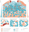
Figure 4. Tumour characteristics that affect the intratumoural fates of nanotheranostics
Slow blood flow may affect the extravasation of nanoparticles in a size-dependent manner. Leaky blood vessels affect the extravasation of nanoparticles in a size-dependent manner. This vasculature leakiness varies considerably between tumours of different types and stages. Dense blood vessels typically enhance tumour accumulation of nanotheranostics. A dense extracellular matrix (ECM), especially in the tumour periphery, may restrict the tumour penetration of nanotheranostics. An increased interstitial fluid pressure (IFP) in many tumours represents a barrier for transcapillary transport of nanotheranostics. Nonspecific uptake by stromal cells, such as tumour-associated macrophages (TAMs), may negatively affect deep tumour penetration and delivery of nanoparticles to cancer cells, but, on the other hand, may serve as an intermediate reservoir.
Figure 5
Figure 5. Nanotheranostics for drug-release monitoring
Designer nanotheranostics have been developed to monitor the intratumoural drug release from nanomedicines by various mechanisms. a | T1 MRI mediators co-released with drug molecules from a liposomal carrier, which generates T1 hyperintensity in magnetic resonance scans. b | Mn2+ and drug molecules (HAsO3)are released on decomposition of Mn2+-doped arsenic trioxide nanoparticles from the mesoporous silica shell, resulting in T1 hyperintensity in magnetic resonance scans. c | T1 mediators are co-released with drug molecules from the polymeric micelle, resulting in T1 hyperintensity in magnetic resonance scans. d | Drugs are released from the surface of iron oxide nanoparticles, resulting in T2 hyperintensity in magnetic resonance scans. e | Iron oxide nanoparticles, Gd-DTPA and drug molecules (5-FU) are loaded in PLGA nanoparticles. On drug release, T1 hyperintensity is generated in the magnetic resonance scans owing to deshielding. f | Chemical-shift agents (for 1H CEST detection) and highly fluorinated compounds (for 19F detection) are loaded into liposomes together with drug molecules. Before release, signal enhancement is generated in 1H CEST magnetic resonance images; after drug release, hyperintensity results in the 19F magnetic resonance images. CEST, chemical exchange-dependent saturation transfer; DTPA, diethylenetriamine pentaacetate; MRI, magnetic resonance imaging; PLGA, poly(lactic-co-glycolic acid); 5-FU, fluorouracil. T1, longitudinal relaxation time; T2, transverse relaxation time.

References

    1. Wilhelm S, et al. Analysis of nanoparticle delivery to tumours. Nat Rev Mater. 2016;1:16014. This perspective stimulates an interesting discussion on the efficiency of nanoparticle delivery to tumours.
    1. Matsumura Y, Maeda H. A new concept for macromolecular therapeutics in cancer chemotherapy: mechanism of tumoritropic accumulation of proteins and the antitumor agent smancs. Cancer Res. 1986;46:6387–6392. - PubMed
    1. Blanco E, Shen H, Ferrari M. Principles of nanoparticle design for overcoming biological barriers to drug delivery. Nat Biotechnol. 2015;33:941–951. - PMC - PubMed
    1. Kobayashi H, Watanabe R, Choyke PL. Improving conventional enhanced permeability and retention (EPR) effects; what is the appropriate target? Theranostics. 2013;4:81–89. - PMC - PubMed
    1. Prabhakar U, et al. Challenges and key considerations of the enhanced permeability and retention effect for nanomedicine drug delivery in oncology. Cancer Res. 2013;73:2412–2417. - PMC - PubMed