. 2017:2017:1238687.
doi: 10.1155/2017/1238687. Epub 2017 Sep 18.

Citalopram Ameliorates Impairments in Spatial Memory and Synaptic Plasticity in Female 3xTgAD Mice

Affiliations

Citalopram Ameliorates Impairments in Spatial Memory and Synaptic Plasticity in Female 3xTgAD Mice

Zhang Wei et al. Biomed Res Int. 2017.

Abstract

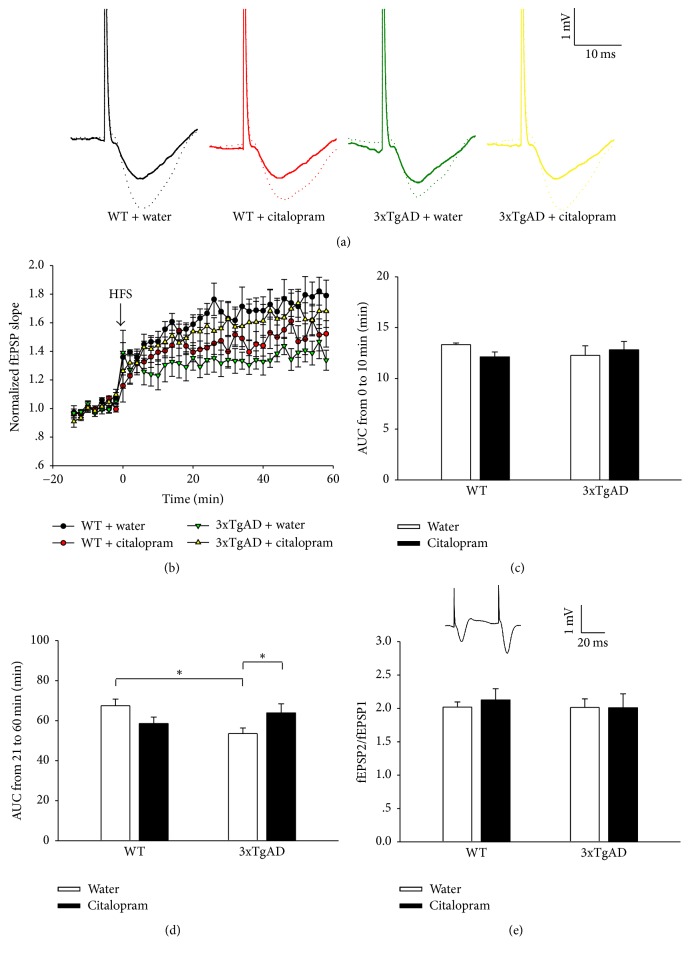
Alzheimer's disease (AD) is the primary cause of dementia. There is no effective treatment. Amyloid-β peptide (Aβ) plays an important role in the pathogenesis and thus strategies suppressing Aβ production and accumulation seem promising. Citalopram is an antidepressant drug and can decrease Aβ production and amyloid plaques in transgenic mice of AD and humans. Whether citalopram can ameliorate memory deficit was not known yet. We tested the effects of citalopram on behavioral performance and synaptic plasticity in female 3xTgAD mice, a well-characterized model of AD. Mice were treated with citalopram or water from 5 months of age for 3 months. Citalopram treatment at approximately 10 mg/kg/day significantly improved spatial memory in the Morris water maze (MWM) test, while not affecting anxiety-like and depression-like behavior in 3xTgAD mice. Further, hippocampal long-term potentiation (LTP) impairment in 3xTgAD mice was reversed by citalopram treatment. Citalopram treatment also significantly decreased the levels of insoluble Aβ40 in hippocampal and cortical tissues in 3xTgAD mice, accompanied with a reduced amyloid precursor protein (APP). Together, citalopram treatment may be a promising strategy for AD and further clinical trials should be conducted to verify the effect of citalopram on cognition in patients with AD or mild cognitive impairment.


Figures

Figure 1
Figure 1
3xTgAD mice showed abnormalities in the open field test at the age of 7-8 months, which were not ameliorated by citalopram. (a) A reduction in locomotor activity was observed in 3xTgAD mice. (b) 3xTgAD mice spent more time in inner square. (c) Representative traces of mice in the open field test. Error bars indicate SEM. P < 0.05, ∗∗P < 0.01, and ∗∗∗P < 0.001.
Figure 2
Figure 2
3xTgAD mice showed abnormalities in the elevated plus maze test and did not present depressive-like behavior in the tail suspension test at the age of 7-8 months. (a) In the elevated plus maze test, a reduction in locomotor activity was observed in 3xTgAD mice, which was not improved by citalopram. (b) In the elevated plus maze test, 3xTgAD mice spent more time in the open arms and citalopram did not take effect. (c) Representative traces of mice in the elevated plus maze test. (d) In the tail suspension test, 3xTgAD mice did not exhibit depressive-like behavior. Error bars indicate SEM. P < 0.05, ∗∗P < 0.01.
Figure 3
Figure 3
Citalopram treatment ameliorated the impairments in spatial learning and memory of 3xTgAD mice in Morris water maze test. (a) Plots showing the changes in the escape latencies of mice in different groups over the 5-day training period. The escape latency of 3xTgAD mice was longer than that of WT mice on the fifth day, and citalopram reversed it. (b) The number of platform crossings was decreased in 3xTgAD mice, which was reversed by citalopram. (c) Representative swimming traces of mice in the probe test. Error bars indicate SEM. P < 0.05, ∗∗P < 0.01.
Figure 4
Figure 4
Citalopram treatment rescued hippocampal LTP of 3xTgAD mice. (a) Sample traces of fEPSPs before HFS (solid line) and 60 min after HFS (dotted line). (b) Time course of fEPSPs and LTP induction before and after HFS. (c) Genotype or citalopram treatment did not affect the induction of LTP. (d) Histograms show that the depressive LTP in 3xTgAD mice was reversed by citalopram. (e) Genotype or citalopram treatment did not affect the PPF (fEPSP2/fEPSP1) in the hippocampal CA1 region. Inset, representative paired fEPSPs. Error bars indicate SEM. P < 0.05.
Figure 5
Figure 5
Citalopram treatment decreased Aβ production by inhibiting APP expression in 3xTgAD mice. (a) The insoluble Aβ40 concentrations in the hippocampus and cerebral cortex samples from citalopram-treated 3xTgAD mice were significantly lower than the concentrations in water-treated 3xTgAD mice. (b) Citalopram treatment did not have significant effect on the levels of insoluble Aβ42 in the hippocampus and cerebral cortex. (c) Representative western blot images of APP and CTFβ in different groups. (d) Citalopram treatment downregulated APP expression in 3xTgAD mice. (e) Citalopram treatment decreased CTFβ levels in 3xTgAD mice. Error bars indicate SEM. P < 0.05, ∗∗P < 0.01, and ∗∗∗P < 0.001.

References

    1. Holtzman D. M., Morris J. C., Goate A. M. Alzheimer's disease: the challenge of the second century. Science Translational Medicine. 2011;3(77) doi: 10.1126/scitranslmed.3002369.77sr1 - DOI - PMC - PubMed
    1. Selkoe D. J. Alzheimer's disease is a synaptic failure. Science. 2002;298(5594):789–791. doi: 10.1126/science.1074069. - DOI - PubMed
    1. Ferreira S. T., Lourenco M. V., Oliveira M. M., De Felice F. G. Soluble amyloid-β oligomers as synaptotoxins leading to cognitive impairment in Alzheimer’s disease. Frontiers in Cellular Neuroscience. 2015;9, article 191 doi: 10.3389/fncel.2015.00191. - DOI - PMC - PubMed
    1. LaFerla F. M., Green K. N., Oddo S. Intracellular amyloid-β in Alzheimer's disease. Nature Reviews Neuroscience. 2007;8(7):499–509. doi: 10.1038/nrn2168. - DOI - PubMed
    1. Mattson M. P. Pathways towards and away from Alzheimer's disease. Nature. 2004;430(7000):631–639. doi: 10.1038/nature02621. - DOI - PMC - PubMed

MeSH terms

LinkOut - more resources