

Estimating CT Image from MRI Data Using 3D Fully Convolutional Networks
- PMID: 29075680
- PMCID: PMC5654583
- DOI: 10.1007/978-3-319-46976-8_18
Estimating CT Image from MRI Data Using 3D Fully Convolutional Networks
Abstract
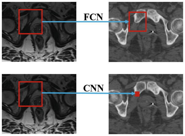
Computed tomography (CT) is critical for various clinical applications, e.g., radiotherapy treatment planning and also PET attenuation correction. However, CT exposes radiation during CT imaging, which may cause side effects to patients. Compared to CT, magnetic resonance imaging (MRI) is much safer and does not involve any radiation. Therefore, recently researchers are greatly motivated to estimate CT image from its corresponding MR image of the same subject for the case of radiotherapy planning. In this paper, we propose a 3D deep learning based method to address this challenging problem. Specifically, a 3D fully convolutional neural network (FCN) is adopted to learn an end-to-end nonlinear mapping from MR image to CT image. Compared to the conventional convolutional neural network (CNN), FCN generates structured output and can better preserve the neighborhood information in the predicted CT image. We have validated our method in a real pelvic CT/MRI dataset. Experimental results show that our method is accurate and robust for predicting CT image from MRI image, and also outperforms three state-of-the-art methods under comparison. In addition, the parameters, such as network depth and activation function, are extensively studied to give an insight for deep learning based regression tasks in our application.
Figures


References
-
- Brenner DJ, Hall EJ. Computed tomographyałan increasing source of radiation exposure. N Engl J Med. 2007;357(22):2277–2284. - PubMed
-
- Burgos N, et al. Robust CT synthesis for radiotherapy planning: application to the head and neck region. In: Navab N, Hornegger J, Wells WM, Frangi AF, editors. MICCAI 2015. LNCS. Vol. 9350. Springer; Heidelberg: 2015. pp. 476–484.
-
- Glorot X, Bengio Y. Understanding the difficulty of training deep feedforward neural networks. International Conference on Artificial Intelligence and Statistics; 2010. pp. 249–256.
Grants and funding
LinkOut - more resources
Full Text Sources
Other Literature Sources