. 2017 Dec;51(6):1674-1684.
doi: 10.3892/ijo.2017.4177. Epub 2017 Oct 23.

Angiotensin receptor blocker telmisartan inhibits cell proliferation and tumor growth of cholangiocarcinoma through cell cycle arrest

Affiliations

Angiotensin receptor blocker telmisartan inhibits cell proliferation and tumor growth of cholangiocarcinoma through cell cycle arrest

Eri Samukawa et al. Int J Oncol. 2017 Dec.

Abstract

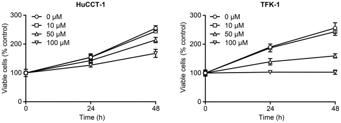
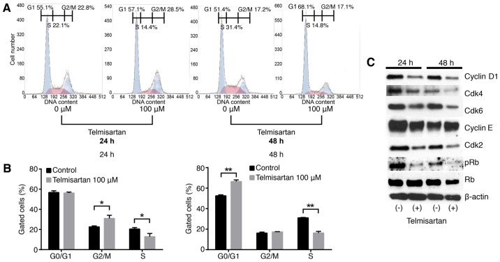
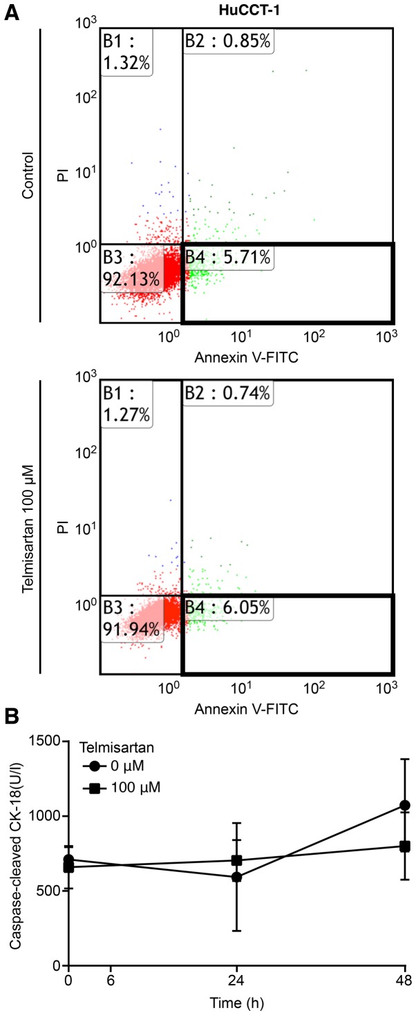
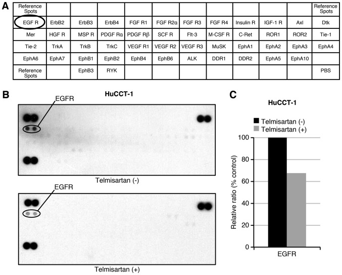
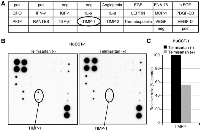
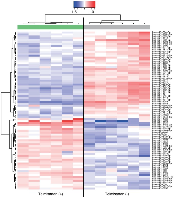
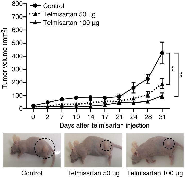
Cholangiocarcinoma (CCA) is at an advanced stage at the time of its diagnosis, and developing a more effective treatment of CCA would be desirable. Angiotensin II type 1 (AT1) receptor blocker (ARB), telmisartan may inhibit cancer cell proliferation, but the mechanisms by which telmisartan affects various cancers remain unknown. In this study, we evaluated the effects of telmisartan on human CCA cells and to assess the expression of microRNAs (miRNAs). We studied the effects of telmisartan on CCA cells using two cell lines, HuCCT-1 and TFK-1. In our experiments, telmisartan inhibited the proliferation of HuCCT-1 and TFK-1 cells. Additionally, telmisartan induced G0/G1 cell cycle arrest via blockade of the G0 to G1 cell cycle transition. Notably, telmisartan did not induce apoptosis in HuCCT-1 cells. This blockade was accompanied by a strong decrease in cell cycle-related protein, especially G1 cyclin, cyclin D1, and its catalytic subumits, Cdk4 and Cdk6. Telmisartan reduced the phosphorylation of EGFR (p-EGFR) and TIMP-1 by using p-RTK and angiogenesis array. Furthermore, miRNA expression was markedly altered by telmisartan in HuCCT-1. Telmisartan inhibits tumor growth in CCA xenograft model in vivo. In conclusion, telmisartan was shown to inhibit human CCA cell proliferation by inducing cell cycle arrest.


Figures

Figure 1
Figure 1
Telmisartan inhibits the proliferation of CCA cells. HuCCT-1 and TFK-1 cells were treated with 0, 10, 50, or 100 µM telmisartan for 24 or 48 h. The data points represent the mean cell number from three independent cultures, and the error bars represent SDs. For each cell line, the conditions at 48 h are significantly different compared with control (0 µM) with P<0.05 as assessed by 2-way ANOVA.
Figure 2
Figure 2
The antiproliferative effects of telmisartan in CCA cells are mediated via cell cycle arrest. (A) HuCCT-1 cells treated with or without 100 µM were analyzed by flow cytometry to estimate the amount of cells in each phase of the cell cycle. (B) The histogram represents the percentage of cells in each cell cycle phase. Telmisartan blocked the cell cycle at G0-G1. (*P<0.05, **P<0.01, vs. control). (C) Expression of cyclin D1, Cdk4, Cdk6, cyclin E, Cdk2, phosphory-lated Rb (pRb) and Rb, in HuCCT-1 cells at 24 and 48 h after the addition of 100 µM telmisartan as analyzed by western blotting.
Figure 3
Figure 3
Telmisartan did not induce apoptosis in HuCCT-1 cells. (A) The early apoptotic changes evoked by 100 µM telmisartan at 24-48 h were assessed by flow cytometry. Annexin V-positive and PI-negative cells were regarded as early apoptotic (enclosed areas in bold squares). Telmisartan did not change the proportion of early apoptotic cancer cells among HuCCT-1 cells. (B) The expression of caspase-cleaved keratin 18 (cCK18), which is produced during apoptosis, was determined using an enzyme-linked immu-nosorbent assay (ELISA). Cells were incubated with or without 100 µM telmisartan.
Figure 4
Figure 4
Effects of telmisartan on p-RTK in HuCCT-1 cells. (A) The template indicates the locations of tyrosine kinase antibodies spotted onto a human phospho-RTK array. (B) Representative expression of various phosphorylated tyrosine kinase receptors in HuCCT-1 cells treated with or without 100 µM telmisartan at 48 h. (C) Densitometry indicated that the ratio of p-EGFR spots in telmisartan-treated compared with untreated cells was 67.4%.
Figure 5
Figure 5
Effects of telmisartan on angiogenesis in HuCCT-1 cells. (A) Template demonstrating the locations of angiogenesis-related proteins spotted onto a human angiogenesis array. (B) Representative expression levels of various angiogenesis-related proteins in HuCCT-1 cells cultured with or without telmisartan. (C) The densitometric ratio of TIMP-1 spots for telmisartan-treated versus untreated cells was 56.0%.
Figure 6
Figure 6
Hierarchical clustering of HuCCT-1 cells cultured with or without telmisartan. The analyzed samples are noted in the columns, and the miRNAs are presented in the rows. The miRNA clustering color scale presented at the top indicates relative miRNA expression levels, with red and blue representing high and low expression levels, respectively.
Figure 7
Figure 7
Telmisartan inhibited the growth of HuCCT-1 cell in a xenograft mouse model. HuCCT-1 cells were subcutaneously implanted into the flanks of nude mice. Then, 50 or 100 µg telmisartan was intraperitoneally injected for 31 days, 5 times per week. The tumors were significantly smaller in telmisartan-treated mice compared with vehicle-treated mice. Each point represents the mean ± standard deviation of 7 animals. P≤0.001 by two-way ANOVA (**P<0.001, vs. control).

Similar articles

Cited by

References

    1. Blechacz B, Gores GJ. Cholangiocarcinoma: Advances in pathogenesis, diagnosis, and treatment. Hepatology. 2008;48:308–321. doi: 10.1002/hep.22310. - DOI - PMC - PubMed
    1. Welzel TM, McGlynn KA, Hsing AW, O'Brien TR, Pfeiffer RM. Impact of classification of hilar cholangiocarcinomas (Klatskin tumors) on the incidence of intra- and extrahepatic cholangiocarcinoma in the United States. J Natl Cancer Inst. 2006;98:873–875. doi: 10.1093/jnci/djj234. - DOI - PubMed
    1. Everhart JE, Ruhl CE. Burden of digestive diseases in the United States Part III: Liver, biliary tract, and pancreas. Gastroenterology. 2009;136:1134–1144. doi: 10.1053/j.gastro.2009.02.038. - DOI - PubMed
    1. Tyson GL, El-Serag HB. Risk factors for cholangiocarcinoma. Hepatology. 2011;54:173–184. doi: 10.1002/hep.24351. - DOI - PMC - PubMed
    1. Kozako T, Soeda S, Yoshimitsu M, Arima N, Kuroki A, Hirata S, Tanaka H, Imakyure O, Tone N, Honda S, et al. Angiotensin II type 1 receptor blocker telmisartan induces apoptosis and autophagy in adult T-cell leukemia cells. FEBS Open Bio. 2016;6:442–460. doi: 10.1002/2211-5463.12055. - DOI - PMC - PubMed

MeSH terms