. 2018 Mar;132(2):449-461.
doi: 10.1007/s00414-017-1714-3. Epub 2017 Oct 26.

A systematic autopsy survey of human infant bridging veins

Affiliations

A systematic autopsy survey of human infant bridging veins

Emma C Cheshire et al. Int J Legal Med. 2018 Mar.

Abstract

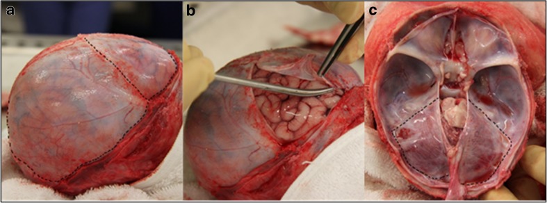
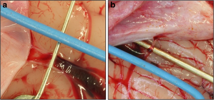
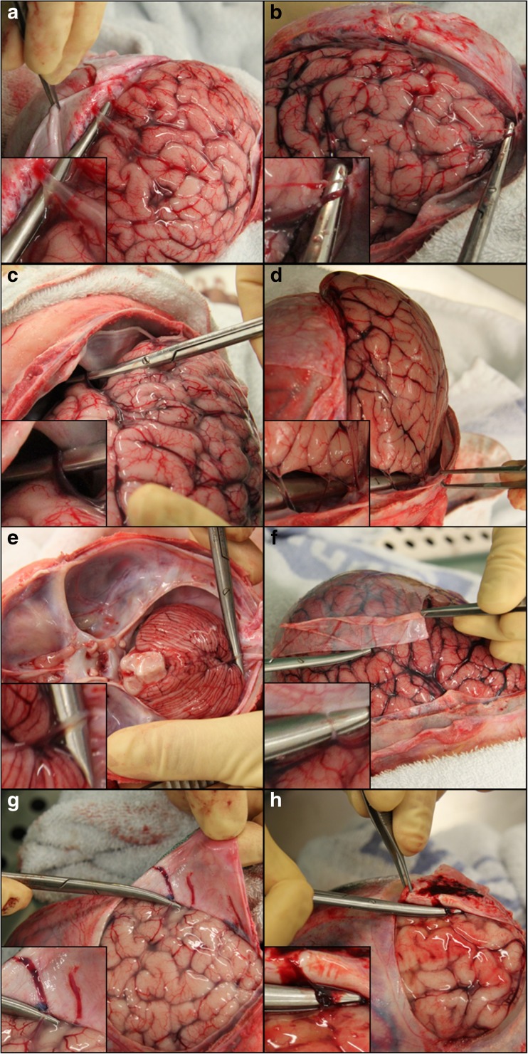
In the first years of life, subdural haemorrhage (SDH) within the cranial cavity can occur through accidental and non-accidental mechanisms as well as from birth-related injury. This type of bleeding is the most common finding in victims of abusive head trauma (AHT). Historically, the most frequent cause of SDHs in infancy is suggested to be traumatic damage to bridging veins traversing from the brain to the dural membrane. However, several alternative hypotheses have been suggested for the cause and origin of subdural bleeding. It has also been suggested by some that bridging veins are too large to rupture through the forces associated with AHT. To date, there have been no systematic anatomical studies on infant bridging veins. During 43 neonatal, infant and young child post-mortem examinations, we have mapped the locations and numbers of bridging veins onto a 3D model of the surface of a representative infant brain. We have also recorded the in situ diameter of 79 bridging veins from two neonatal, one infant and two young children at post-mortem examination. Large numbers of veins, both distant from and directly entering the dural venous sinuses, were discovered travelling between the brain and dural membrane, with the mean number of veins per brain being 54.1 and the largest number recorded as 94. The mean diameter of the bridging veins was 0.93 mm, with measurements ranging from 0.05 to 3.07 mm. These data demonstrate that some veins are extremely small and subjectively, and they appear to be delicate. Characterisation of infant bridging veins will contribute to the current understanding of potential vascular sources of subdural bleeding and could also be used to further develop computational models of infant head injury.

Keywords: Abuse; Bridging vein; Child; Head injury; Post-mortem; Subdural haemorrhage.


Conflict of interest statement

Ethical approval

All procedures performed in studies involving human participants were in accordance with the ethical standards of the institutional and/or national research committee and with the 1964 Helsinki declaration and its later amendments or comparable ethical standards.

Retrospective ethical approval was given for the use of anonymised archived autopsy images for the purpose of 3D anatomical mapping of bridging veins. For cases involving the use of digital photography for vein measuring purposes, and MRI scanning, appropriate parental consent was prospectively obtained.

Conflict of interest

RDGM regularly provides expert opinion to various Courts in relation to paediatric head injury and receives payment for doing so. Otherwise, the authors declare that they have no conflict of interest.

Figures

Fig. 1
Fig. 1
Incising and reflecting the dural membrane. a Incision lines along the right coronal and lambdoid sutures. b Reflection of the right frontal dura mater. c Incision lines on the tentorium
Fig. 2
Fig. 2
Digital macroscopic photographs of bridging veins with blue wire of known diameter. a Bridging vein on the left hemisphere in the frontal parasagittal region. b Bridging vein on the superior surface of the cerebellum
Fig. 3
Fig. 3
Reflection of the dura mater to reveal the bridging veins with ×2–4 magnification insets on the bottom left hand corner of each image. a Parasagittal bridging vein entering the SSS. b Bridging veins at the frontal pole. c Temporal pole. d Inferior aspect of the occipital lobe. e Bridging vein on the superior surface of the cerebellum. f Bridging vein distant from the sinuses, on the right parietal convexity. g Bridging veins running towards the SSS on the inner aspect of the dura. h AHT case with bridging vein engorged with blood and not blanching under slight pressure
Fig. 4
Fig. 4
Parasagittal and tentorial sinuses. a Two bridging veins (arrowheads) draining the frontal lobe and entering a parasagittal sinus (arrow) approximately 3 cm distal to the SSS. b Parasagittal sinus from image a after cutting through the bridging veins and further reflection of the dura, arrowheads show the locations where the two bridging veins joined the sinus. c Tentorial sinuses (arrows) within the dura on both hemispheres of the cerebellum
Fig. 5
Fig. 5
Heat map distribution of infant bridging veins. a Superior view showing interhemispheric fissure. b Inferior view showing vein distribution on the inferior temporal, occipital and frontal lobe. c Left lateral view. d Right lateral view. e Posterior view. f Anterior view showing veins near the frontal pole. g Interhemispheric fissure, right hemisphere. h Interhemispheric fissure, left hemisphere. i Superior view of the cerebellum. j Heat map intensity bar
Fig. 6
Fig. 6
Dot scatter plots of infant bridging veins. a Superior view showing interhemispheric fissure. b Inferior view showing veins on the inferior temporal, occipital and frontal lobe. c Left lateral view. d Right lateral view. e Posterior view. f Anterior view showing veins near the frontal pole. g Interhemispheric fissure, right hemisphere. h Interhemispheric fissure, left hemisphere. i Superior view of the cerebellum

References

    1. Feldman KW, Bethel R, Shugerman RP, Grossman DC, Grady MS, Ellenbogen RG. The cause of infant and toddler subdural haemorrhage: a prospective study. Pediatr. 2001;108:636–646. doi: 10.1542/peds.108.3.636. - DOI - PubMed
    1. Hoskote A, Richards P, Anslow P, McShane T. Subdural haematoma and non-accidental head injury in children. Childs Nerv Syst. 2002;18:311–317. doi: 10.1007/s00381-002-0616-x. - DOI - PubMed
    1. Tzioumi D, Oates RK. Subdural hematomas in children under 2 years. Accidental or inflicted? A 10-year experience. Child Abuse Negl. 1998;22:1105–1112. doi: 10.1016/S0145-2134(98)00093-3. - DOI - PubMed
    1. Hobbs C, Childs AM, Wynne J, Livingston J, Seal A. Subdural haematoma and effusion in infancy: an epidemiological study. Arch Dis Child. 2005;90:952–955. doi: 10.1136/adc.2003.037739. - DOI - PMC - PubMed
    1. Rooks VJ, Eaton JP, Ruess L, Petermann GW, Keck-Wherley J, Pedersen RC. Prevalence and evolution of intracranial hemorrhage in asymptomatic term infants. Am J Neuroradiol. 2008;29:1082–1089. doi: 10.3174/ajnr.A1004. - DOI - PMC - PubMed

Grants and funding