Review
. 2018 Apr;21(3):261-268.
doi: 10.1111/ner.12706. Epub 2017 Oct 27.

High-Resolution Multi-Scale Computational Model for Non-Invasive Cervical Vagus Nerve Stimulation

Affiliations
Review

High-Resolution Multi-Scale Computational Model for Non-Invasive Cervical Vagus Nerve Stimulation

Antonios P Mourdoukoutas et al. Neuromodulation. 2018 Apr.

Abstract

Objectives: To develop the first high-resolution, multi-scale model of cervical non-invasive vagus nerve stimulation (nVNS) and to predict vagus fiber type activation, given clinically relevant rheobase thresholds.

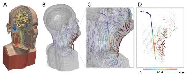
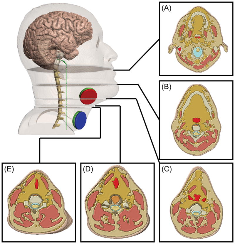
Methods: An MRI-derived Finite Element Method (FEM) model was developed to accurately simulate key macroscopic (e.g., skin, soft tissue, muscle) and mesoscopic (cervical enlargement, vertebral arch and foramen, cerebral spinal fluid [CSF], nerve sheath) tissue components to predict extracellular potential, electric field (E-Field), and activating function along the vagus nerve. Microscopic scale biophysical models of axons were developed to compare axons of varying size (Aα-, Aβ- and Aδ-, B-, and C-fibers). Rheobase threshold estimates were based on a step function waveform.

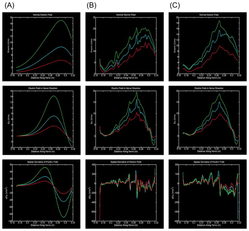
Results: Macro-scale accuracy was found to determine E-Field magnitudes around the vagus nerve, while meso-scale precision determined E-field changes (activating function). Mesoscopic anatomical details that capture vagus nerve passage through a changing tissue environment (e.g., bone to soft tissue) profoundly enhanced predicted axon sensitivity while encapsulation in homogenous tissue (e.g., nerve sheath) dulled axon sensitivity to nVNS.

Conclusions: These findings indicate that realistic and precise modeling at both macroscopic and mesoscopic scales are needed for quantitative predictions of vagus nerve activation. Based on this approach, we predict conventional cervical nVNS protocols can activate A- and B- but not C-fibers. Our state-of-the-art implementation across scales is equally valuable for models of spinal cord stimulation, cortex/deep brain stimulation, and other peripheral/cranial nerve models.

Keywords: Cranial nerve stimulation; electrode placement; mechanisms of action; neurostimulation; stimulation; vagus nerve stimulation.


Conflict of interest statement

Conflict of Interest: Dr. Simon is an employee of electroCore and has shares in the company. The City University of New York has patents on brain stimulation with Dr. Bikson as inventor. Dr. Bikson also has equity in Soterix Medical Inc. and serves as a scientific advisor to Boston Scientific Inc.

Figures

Figure 1
Figure 1. High-resolution model of nVNS current flow
(A) MRI derived model including bone, brain, muscle and other soft tissue masks, and vagus nerve (green). (B) Stimulation of nVNS with electrode placement showing flux lines map gross current flow patterns through neck, with false color of local current density (>10 A/m2 max). Gross current flow patterns are determined by electrode position and anatomy. (C) Inset showing expansion of current flow around vagus nerve (1.44 A/m2 max) using the given electrode montage. (D) Arrow plots of gross current density pattern and current density on vagus nerve in false colors. The current density (proportional to electric field) along the nerve supports the prediction of activation, depending on fiber type. All models are under the quasi-static assumption with the anode in red and cathode in blue for illustration of instant direction.
Figure 2
Figure 2. Role of tissue properties around nerve in predicting driving forces for activation
Three drivers of neuronal polarization: Electric field magnitude (top row), electric field component aligned with the nerve (middle row), and spatial derivative of electric field (activating function) in the direction of the nerve (bottom row). Three conditions of tissue detail are modeled: (A, first column) simplified homogenous soft tissue (muscle, fat, ligament, intervertebral disk merged); (B, second column) full inhomogeneous soft tissue anatomy without a fat sheath surrounding the vagus nerve; (C, third column) full inhomogenous soft tissue anatomy with a fat sheath surrounding the vagus nerve. In each case, soft tissue conductivity was doubled (red), unaffected (blue) and halved (green). Whereas in a simplified homogeneous soft tissue case (A) drivers of activation are smooth, with full inhomogeneous soft tissue anatomy (B, C) local maximum are observed (0.217 m, 0.250 m and 0.269 m) corresponding to changing tissue around the nerve (see Figure 4). The addition of a sheath generally dulls the influence of these transitions.
Figure 3
Figure 3. Illustration of local tissue inhomogeneity around nerve leading to transients in drivers of activation
(A) 0.180 m (B) 0.217 m (C) 0.250 m (D) 0.263 m (E) 0.269 m. Five anatomical cross sections showing cases in which either fat and soft tissue (A, B, C, E) or just fat (D) borders the vagus nerve. Slices (B, C, D, E) are relatively close to the stimulating electrodes while slice (A) is relatively far.

References

    1. Tracey KJ. Physiology and immunology of the cholinergic antiinflammatory pathway. The Journal of clinical investigation. 2007;117(2):289–296. - PMC - PubMed
    1. Bonaz B, Picq C, Sinniger V, Mayol J-F, Clarençon D. Vagus nerve stimulation: from epilepsy to the cholinergic anti-inflammatory pathway. Neurogastroenterol Motil. 2013;25(3):208–221. - PubMed
    1. Zhou L, Lin J, Lin J, Kui G, Zhang J, Yu Y. Neuroprotective effects of vagus nerve stimulation on traumatic brain injury. Neural regeneration research. 2014;9(17):1585. - PMC - PubMed
    1. Hays SA, Rennaker RL, Kilgard MP. Targeting plasticity with vagus nerve stimulation to treat neurological disease. Progress in brain research. 2013;207:275. - PMC - PubMed
    1. Van Leusden JW, Sellaro R, Colzato LS. Transcutaneous Vagal Nerve Stimulation (tVNS): a new neuromodulation tool in healthy humans? Front Psychol. 2015;6:102. - PMC - PubMed