. 2018 Jun 11;57(24):7171-7175.
doi: 10.1002/anie.201709184. Epub 2018 Apr 25.

G-Quadruplex Secondary Structure Obtained from Circular Dichroism Spectroscopy

Affiliations

G-Quadruplex Secondary Structure Obtained from Circular Dichroism Spectroscopy

Rafael Del Villar-Guerra et al. Angew Chem Int Ed Engl. .

Abstract

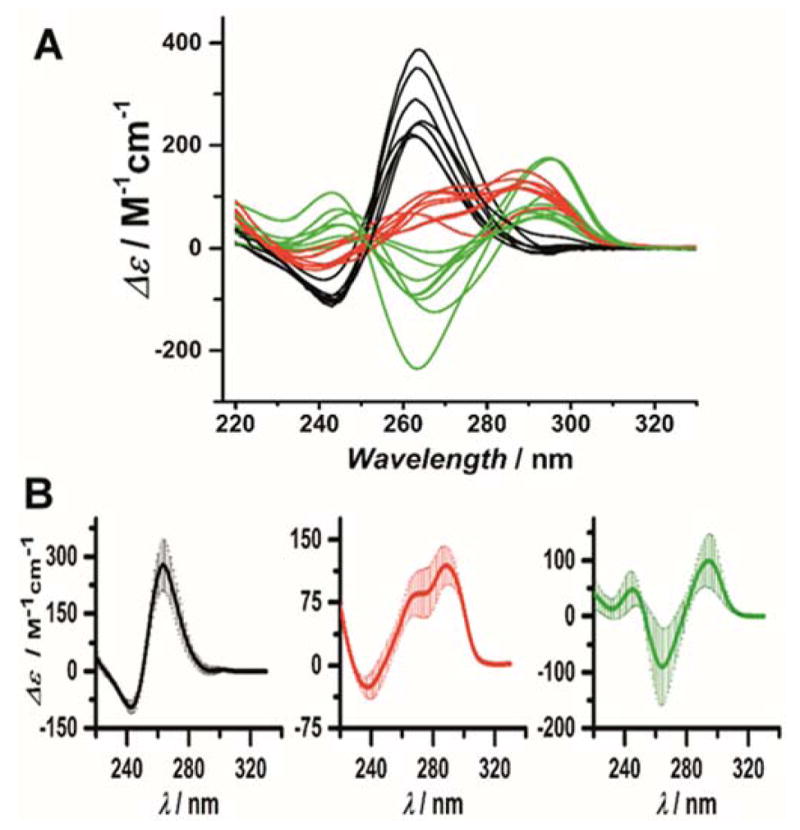
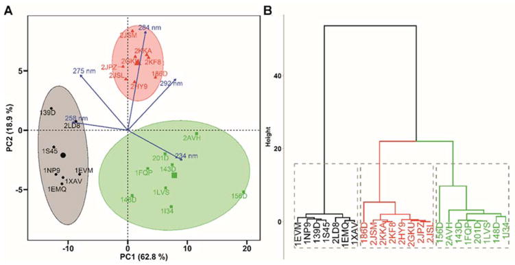
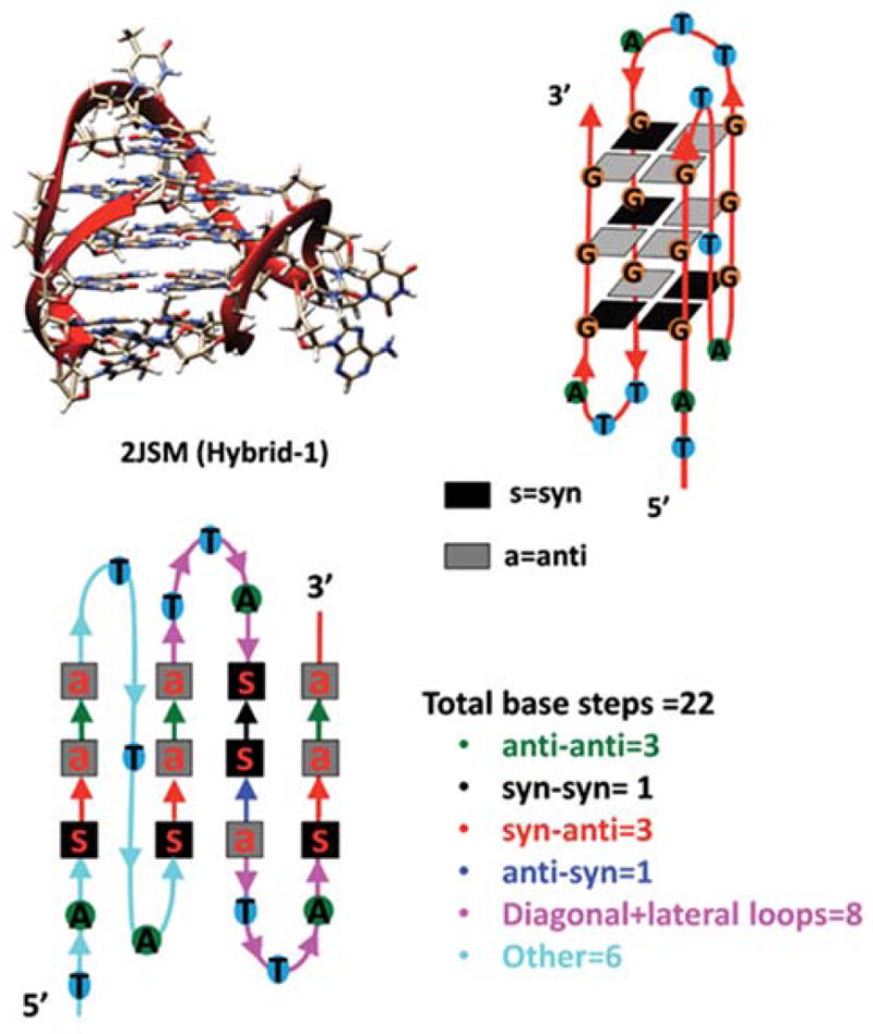
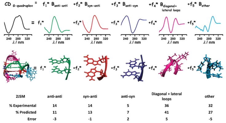
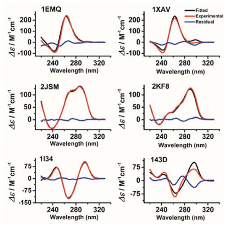
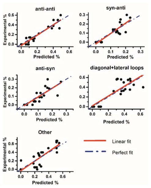
A curated library of circular dichroism spectra of 23 G-quadruplexes of known structure was built and analyzed. The goal of this study was to use this reference library to develop an algorithm to derive quantitative estimates of the secondary structure content of quadruplexes from their experimental CD spectra. Principal component analysis and singular value decomposition were used to characterize the reference spectral library. CD spectra were successfully fit to obtain estimates of the amounts of base steps in anti-anti, syn-anti or anti-syn conformations, in diagonal or lateral loops, or in other conformations. The results show that CD spectra of nucleic acids can be analyzed to obtain quantitative structural information about secondary structure content in an analogous way to methods used to analyze protein CD spectra.

Keywords: DNA; G-quadruplexes; circular dichroism; secondary structure.


Figures

Figure 1
Figure 1
A) CD spectra of the reference library of 23 G-quadruplex. B) Average CD spectra obtained for cluster 1(left, black), cluster 2 (center, red) and cluster 3 (right, green). The associated standard deviations are represented by the shaded areas.
Figure 2
Figure 2
(A) Principal component analysis (PCA) and (B) Hierarchical Clustering on Principal Components (HCPC) of the reference CD spectra library of G-quadruplexes. In panel (A), the score plot is shown in which the first and second principal components are plotted against one another. The blue arrows indicate the loading vectors that drive the clustering.
Figure 3
Figure 3
Schematic illustration of the definition of the G-quadruplex secondary structure elements used in this study. The example shown is for the human telomere hybrid 1 structure with the PDB identifier 2JSM.
Figure 4
Figure 4
Schematic illustration of the method used to calculate the fractions of different secondary structural elements by constrained least-square fitting of a test CD spectrum to the five secondary basis spectra.
Figure 5
Figure 5
Experimental (red), fitted (black) and residual (blue) CD spectra for selected G-quadruplexes obtained by nonlinear least-squares fitting to eq. 1. The PDB identifier for each structure is shown in each panel.
Figure 6
Figure 6
Scatter plots of the fractions of secondary structural elements determined from the known structures (“Experimental %”) versus the fractions obtained by fits to experimental CD spectra (“Predicted %). These data were obtained using the reference G-quadruplex CD spectra library and a leave-one-out cross-validation constrained least-squares fitting strategy. The red solid line is the least-squares linear fit to the data points, while the dashed blue line represents the line (slope =1) expected for a perfect correlation between the actual and estimated fractions. The correlation coefficients for the linear fits are shown in Table S6.

References

    1. Hansel-Hertsch R, Di Antonio M, Balasubramanian S. Nat Rev Mol Cell Biol. 2017;18:279–284. - PubMed
    1. Kypr J, Kejnovská I, Renčiuk D, Vorlíčková M. Nucleic Acids Research. 2009;37:1713–1725. - PMC - PubMed
    2. Vorlíčková M, Kejnovská I, Bednářová K, Renčiuk D, Kypr J. Chirality. 2012;24:691–698. - PubMed
    1. Randazzo A, Spada G, da Silva M. Top Curr Chem. 2013;330:67–86. - PubMed
    2. Vorlíčková M, Kejnovská I, Sagi J, Renčiuk D, Bednářová K, Motlová J, Kypr J. Methods. 2012;57:64–75. - PubMed
    3. Karsisiotis AI, Hessari NM, Novellino E, Spada GP, Randazzo A, Webba da Silva M. Angewandte Chemie, International Edition. 2011;50:10645. - PubMed
    4. Masiero S, Trotta R, Pieraccini S, De Tito S, Perone R, Randazzo A, Spada GP. Organic & Biomolecular Chemistry. 2010;8:2683–2692. - PubMed
    5. Paramasivan S, Rujan I, Bolton PH. Methods. 2007;43:324–331. - PubMed
    1. Jaumot J, Eritja R, Navea S, Gargallo R. Analytica Chimica Acta. 2009;642:117–126. - PubMed
    1. del Villar-Guerra R, Gray RD, Chaires JB. Curr Protoc Nucleic Acid Chem. 2017;68:17.8.1–17.8.16. - PMC - PubMed

Publication types