Clinical Significance of Prostatic Calculi: A Review
- PMID: 29076299
- PMCID: PMC5756803
- DOI: 10.5534/wjmh.17018
Clinical Significance of Prostatic Calculi: A Review
Abstract
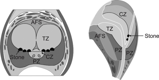
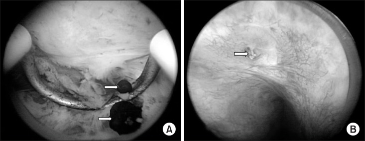
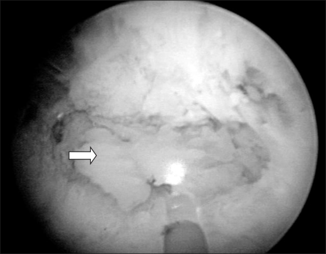
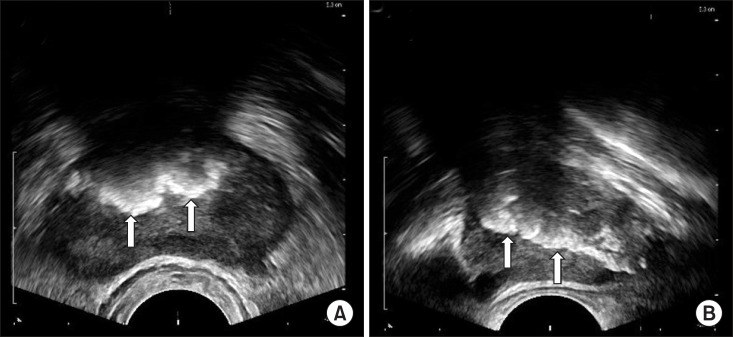
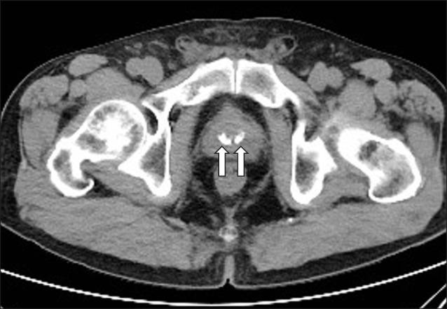
Prostatic calculi often occur in middle-aged and old men. Prostatic calculi are usually classified as primary/endogenous stones or secondary/extrinsic stones. Endogenous stones are commonly caused by obstruction of the prostatic ducts around the enlarged prostate by benign prostatic hyperplasia (BPH) or by chronic inflammation. Extrinsic stones occur mainly around the urethra, because they are caused by urine reflux. The exact prevalence of prostatic calculi is not known, and it has been reported to vary widely, from 7% to 70%. Most cases of prostatic calculi are not accompanied by symptoms. Therefore, most cases are found incidentally during the diagnosis of BPH using transrectal ultrasonography (TRUS). However, prostatic calculi associated with chronic prostatitis may be accompanied by chronic pelvic pain. Rare cases have been reported in which extrinsic prostatic calculi caused by urine reflux have led to voiding difficulty due to their size. More than 80% of prostatic calculi are composed of calcium phosphate. Prostatic calculi can be easily diagnosed using TRUS or computed tomography. Treatment is often unnecessary, but if an individual experiences difficulty in urination or chronic pain, prostatic calculi can be easily removed using a transurethral electroresection loop or holmium laser.
Keywords: Calculi; Prostate; Prostatic hyperplasia; Prostatitis.
Copyright © 2018 Korean Society for Sexual Medicine and Andrology.
Conflict of interest statement
The author has no potential conflicts of interest to disclose.
Figures
References
-
- Klimas R, Bennett B, Gardner WA., Jr Prostatic calculi: a review. Prostate. 1985;7:91–96. - PubMed
-
- Lee CH, Akin-Olugbade O, Kirschenbaum A. Overview of prostate anatomy, histology, and pathology. Endocrinol Metab Clin North Am. 2011;40:565–575. - PubMed
-
- Selman SH. The McNeal prostate: a review. Urology. 2011;78:1224–1228. - PubMed
-
- Young HH. Prostatic calculi. J Urol. 1934;32:660–709.
-
- McDonald HP, Upchurch WE, Sturdevant CE. Treatment of prostatic calculi. JAMA. 1955;157:787–788. - PubMed
Publication types
LinkOut - more resources
Full Text Sources
Other Literature Sources