Review
. 2018 Jan;28(1):9-21.
doi: 10.1038/cr.2017.133. Epub 2017 Oct 27.

Plasma membrane changes during programmed cell deaths

Affiliations
Review

Plasma membrane changes during programmed cell deaths

Yingying Zhang et al. Cell Res. 2018 Jan.

Abstract

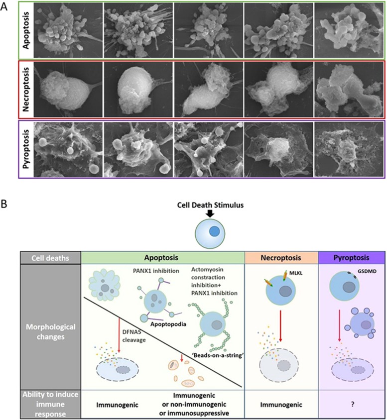
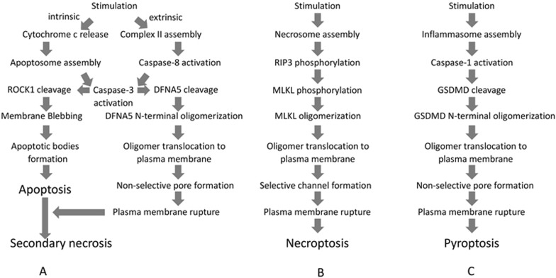
Ruptured and intact plasma membranes are classically considered as hallmarks of necrotic and apoptotic cell death, respectively. As such, apoptosis is usually considered a non-inflammatory process while necrosis triggers inflammation. Recent studies on necroptosis and pyroptosis, two types of programmed necrosis, revealed that plasma membrane rupture is mediated by MLKL channels during necroptosis but depends on non-selective gasdermin D (GSDMD) pores during pyroptosis. Importantly, the morphology of dying cells executed by MLKL channels can be distinguished from that executed by GSDMD pores. Interestingly, it was found recently that secondary necrosis of apoptotic cells, a previously believed non-regulated form of cell lysis that occurs after apoptosis, can be programmed and executed by plasma membrane pore formation like that of pyroptosis. In addition, pyroptosis is associated with pyroptotic bodies, which have some similarities to apoptotic bodies. Therefore, different cell death programs induce distinctive reshuffling processes of the plasma membrane. Given the fact that the nature of released intracellular contents plays a crucial role in dying/dead cell-induced immunogenicity, not only membrane rupture or integrity but also the nature of plasma membrane breakdown would determine the fate of a cell as well as its ability to elicit an immune response. In this review, we will discuss recent advances in the field of apoptosis, necroptosis and pyroptosis, with an emphasis on the mechanisms underlying plasma membrane changes observed on dying cells and their implication in cell death-elicited immunogenicity.


Figures

Figure 1
Figure 1
Morphological features of apoptosis, necroptosis, and pyroptosis and their linkages with immunogenicity. (A) Dying cells revealed by scanning electron microscopy. In RAW264.7 cells, apoptosis was induced by TNF+Smac mimetics; necroptosis was induced by TNF+Smac mimetics+zVAD; pyroptosis was induced by LPS priming followed by nigericin treatment. (B) Membrane blebbing followed by formation of apoptotic bodies is commonly observed in apoptosis. Under certain conditions, such as inhibition of PANX1 by trovafloxacin or further combined inhibition of actomyosin contraction by cytochalasin D or GSK 269962, apoptotic cells exhibit two apoptotic body-related morphological changes called apoptopodia and 'beads-on-a-string' protrusions. These membrane-enveloped fragments can be immunogenic, non-immunogenic, or even immunosuppressive under different experimental settings. However, the regulated secondary necrosis of apoptotic cells mediated by DFNA5 can be highly inflammatory. In necroptosis, MLKL-mediated plasma membrane rupture leads to release of cellular contents and thus immunogenicity. Pyroptosis results from an inflammatory response induced by inflammasome activation, which is frequently observed in professional phagocytes and tightly associated with IL-1β/IL-18 secretion. Whether GSDMD-mediated pyroptosis itself is immunogenic awaits further investigation.
Figure 2
Figure 2
Outlines of the signal transduction pathways leading to plasma membrane changes in apoptosis (including secondary necrosis), necroptosis, and pyroptosis. (A) Apoptosis can be initiated by either intrinsic or extrinsic pathway. Caspase-3 activation resulting from either pathway cleaves ROCK1 to promote plasma membrane blebbing, followed by generation of apoptotic bodies. Caspase-3 can also cleave DFNA5 to generate the DFNA5 N-terminal fragment, which forms oligomers and translocates to the plasma membrane, leading to its rupture by the formation of non-selective pores and finally secondary necrosis. (B) In the necroptotic pathway, various external death ligands can initiate necrosome assembly. Once in the necrosome, RIP3 is autophosphorylated. Phosphorylated RIP3 recruits and phosphorylates MLKL, leading to MLKL oligomerization and translocation to the plasma membrane. MLKL oligomers execute necroptosis by generating cation channels, causing plasma membrane rupture. (C) Pyroptotic stimulation elicits inflammasome formation and subsequent caspase-1 activation. Activated caspase-1 cleaves GSDMD, generating the GSDMD N-terminal fragment, which oligomerizes and translocates to the plasma membrane and causes plasma membrane rupture via non-selective pore formation.

References

    1. Walker NI, Harmon BV, Gobe GC, et al. Patterns of cell death. Methods Achiev Exp Pathol 1988; 13:18–54. - PubMed
    1. Nagata S, Tanaka M. Programmed cell death and the immune system. Nat Rev Immunol 2017; 17:333–340. - PubMed
    1. Rogers C, Fernandes-Alnemri T, Mayes L, et al. Cleavage of DFNA5 by caspase-3 during apoptosis mediates progression to secondary necrotic/pyroptotic cell death. Nat Commun 2017; 8:14128. - PMC - PubMed
    1. Kono H, Rock KL. How dying cells alert the immune system to danger. Nat Rev Immunol 2008; 8:279–289. - PMC - PubMed
    1. Wallach D, Kang TB, Dillon CP, et al. Programmed necrosis in inflammation: toward identification of the effector molecules. Science 2016; 352:aaf2154. - PubMed

Publication types