. 2017 Oct 27;9(11):346.
doi: 10.3390/toxins9110346.

The Aromatic Head Group of Spider Toxin Polyamines Influences Toxicity to Cancer Cells

Affiliations

The Aromatic Head Group of Spider Toxin Polyamines Influences Toxicity to Cancer Cells

David Wilson et al. Toxins (Basel). .

Abstract

Spider venoms constitute incredibly diverse libraries of compounds, many of which are involved in prey capture and defence. Polyamines are often prevalent in the venom and target ionotropic glutamate receptors. Here we show that a novel spider polyamine, PA366, containing a hydroxyphenyl-based structure is present in the venom of several species of tarantula, and has selective toxicity against MCF-7 breast cancer cells. By contrast, a polyamine from an Australian funnel-web spider venom, which contains an identical polyamine tail to PA366 but an indole-based head-group, is only cytotoxic at high concentrations. Our results suggest that the ring structure plays a role in the cytotoxicity and that modification to the polyamine head group might lead to more potent and selective compounds with potential as novel cancer treatments.

Keywords: NMR spectroscopy; cancer; cytotoxicity; polyamine; spider venom.


Conflict of interest statement

The authors declare no conflict of interest.

Figures
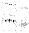
Figure 1
Figure 1
Cytotoxicity of Phlogius sp. crude venom and a purified component, PA366. (A) Dose response for cytotoxic cell killing by Phlogius sp. crude venom compared to vehicle. Crude venom (squares) or vehicle (circles) was incubated with MCF-7 breast cancer cells at the indicated volumes for 5 days, before assay for cell survival using the sulforhodamine B (SRB) assay. Cell survival percentages normalised to untreated cells are indicated. (B) Dose response for cytotoxic cell death of PA366 compared to vehicle in MCF-7, SK-MEL-28 or NFF cells. Cells were treated with the indicated concentrations of purified PA366 for 5 days, before assay for cell survival using SRB. Cell survival percentages normalised to untreated cells are indicated. Representative data from a single experiment with triplicate readings are shown.
Figure 2
Figure 2
Characterisation of spider venom polyamines. (A) RP-HPLC chromatogram of crude female Phlogius sp. venom with the peak corresponding to the polyamine PA366 highlighted in red (Thermo Scientific Hypersil GOLD aQ 250 × 10 mm, 5 µm column; 1 mL/min flow rate; Solvent A H2O/0.05% TFA, Solvent B 90% ACN/H2O/0.045% TFA; 5–80% solvent B in 75 min, 80–90% solvent B in 5 min, 90% solvent B for 5 min, and 90–5% solvent B in 2 min; absorbance at 214 nm); (B) SCIEX TOF/TOF™ 5800 MALDI MS/MS spectrum of PA366 using CHCA matrix, and the determined chemical structure with relevant fragment ions highlighted; (C) RP-HPLC chromatogram of crude female A. robustus venom with the peak corresponding to the polyamine PA389 highlighted in red (Thermo Scientific Hypersil GOLD aQ 250 × 10 mm, 5 µm column; 1 mL/min flow rate; Solvent A H2O/0.05% TFA, Solvent B 90% ACN/H2O/0.045% TFA; 5–80% solvent B in 75 min, 80–90% solvent B in 5 min, 90% solvent B for 5 min, and 90–5% solvent B in 2 min; absorbance at 214 nm); (D) SCIEX TOF/TOF™ 5800 MALDI-MS/MS spectrum of PA389 using CHCA matrix, and the determined chemical structure with relevant fragment ions highlighted.
Figure 3
Figure 3
Chemical structure and 1H NMR spectrum of PA366. The assignments were derived based on two-dimensional NMR spectra and confirmed using mass spectrometry fragmentation analysis.
Figure 4
Figure 4
One-dimensional NMR spectra of selected crude spider venoms and purified polyamines. 1H NMR spectra of crude venom from Phlogius sp., A. robustus and C. darlingi recorded at 600 MHz, showing the presence of PA366 in the Phlogius sp. venom, and PA389 in the A. robustus and C. darlingi venom. The 1H NMR spectra of purified PA366 and PA389 recorded at 600 MHz are also shown.
Figure 5
Figure 5
Cytotoxicity of PA389 against cancer cell lines. Dose response for cytotoxic cell killing of PA389 in MCF-7 (circles) or SK-MEL-28 (squares) cells. Cells were treated with the indicated concentrations of purified PA389 for five days, before assay for cell survival was assessed using SRB. Cell survival percentages normalised to untreated cells are indicated. Representative data from a single experiment with triplicate readings are shown.
Figure 6
Figure 6
One-dimensional NMR spectra of selected crude spider venoms from P. antinous and A. geniculata. 1H NMR analysis of the crude venom from P. antinous shows a composition that is primarily peptides, based on the number of peaks and dispersion in the amide region; this venom does not show any evidence of cytotoxicity against MCF-7, SK-MEL-28 or NFF cells. In contrast, the spectrum of cytotoxic crude venom from A. geniculata is dominated by the peaks corresponding to PA366.
Figure 7
Figure 7
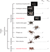
Phylogenic tree of some spider venoms showing the distribution of PA366 and PA389. A phylogenetic tree of crude spider venoms that demonstrated cytotoxicity on MCF-7 or SK-MEL-28 cells in this study, and the presence of the polyamines PA366 and PA389. The two spider species, Hebestatis theveneti and Harpactirella sp., from the original identification and characterisation study of PA389 are also included [19] (Photograph credits: Acanthoscurria geniculata, Ceratogyrus darlingi, Harpactirella sp., Hysterocrates gigas, and Nhandu chromatus—Bastian Rast; Chilobrachys penang—Muhammad Ashraf; Psalmopoeus irminia—Edward Evans; Hebestatis theveneti—Marshal Hedin; Phlogius sp. and Atrax robustus—David Wilson).

References

    1. Deyrup S.T., Eckman L.E., McCarthy P.H., Smedley S.R., Meinwald J., Schroeder F.C. 2D NMR-spectroscopic screening reveals polyketides in ladybugs. Proc. Natl. Acad. Sci. USA. 2011;108:9753–9758. doi: 10.1073/pnas.1107020108. - DOI - PMC - PubMed
    1. Shanmugam M.K., Lee J.H., Chai E.Z., Kanchi M.M., Kar S., Arfuso F., Dharmarajan A., Kumar A.P., Ramar P.S., Looi C.Y., et al. Cancer prevention and therapy through the modulation of transcription factors by bioactive natural compounds. Semin. Cancer Biol. 2016;40–41:35–47. doi: 10.1016/j.semcancer.2016.03.005. - DOI - PubMed
    1. Doroghazi J.R., Albright J.C., Goering A.W., Ju K.S., Haines R.R., Tchalukov K.A., Labeda D.P., Kelleher N.L., Metcalf W.W. A roadmap for natural product discovery based on large-scale genomics and metabolomics. Nat. Chem. Biol. 2014;10:963–968. doi: 10.1038/nchembio.1659. - DOI - PMC - PubMed
    1. Wickenden A., Priest B., Erdemli G. Ion channel drug discovery: Challenges and future directions. Future Med. Chem. 2012;4:661–679. doi: 10.4155/fmc.12.4. - DOI - PubMed
    1. Escoubas P., Sollod B., King G.F. Venom landscapes: Mining the complexity of spider venoms via a combined cdna and mass spectrometric approach. Toxicon. 2006;47:650–663. doi: 10.1016/j.toxicon.2006.01.018. - DOI - PubMed

Publication types

LinkOut - more resources