. 2017 Oct 27;18(11):2259.
doi: 10.3390/ijms18112259.

Development of A Chimeric Antigen Receptor Targeting C-Type Lectin-Like Molecule-1 for Human Acute Myeloid Leukemia

Affiliations

Development of A Chimeric Antigen Receptor Targeting C-Type Lectin-Like Molecule-1 for Human Acute Myeloid Leukemia

Eduardo Laborda et al. Int J Mol Sci. .

Abstract

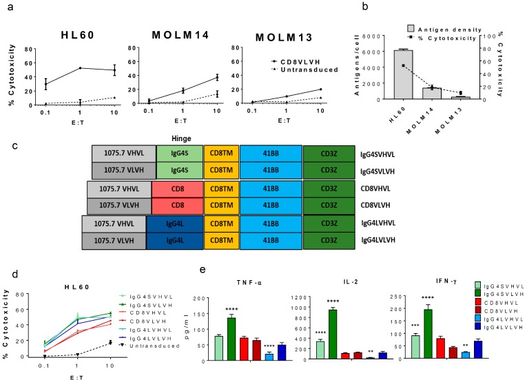
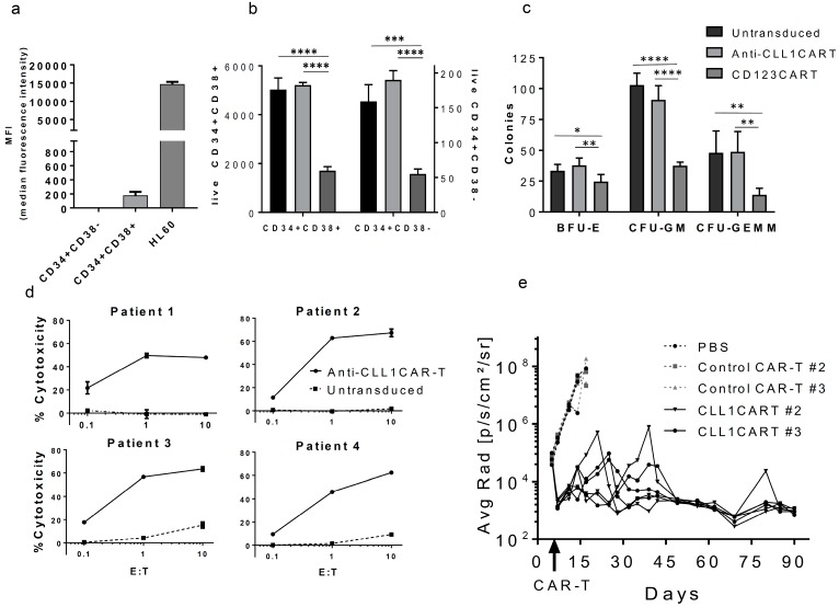
The treatment of patients with acute myeloid leukemia (AML) with targeted immunotherapy is challenged by the heterogeneity of the disease and a lack of tumor-exclusive antigens. Conventional immunotherapy targets for AML such as CD33 and CD123 have been proposed as targets for chimeric antigen receptor (CAR)-engineered T-cells (CAR-T-cells), a therapy that has been highly successful in the treatment of B-cell leukemia and lymphoma. However, CD33 and CD123 are present on hematopoietic stem cells, and targeting with CAR-T-cells has the potential to elicit long-term myelosuppression. C-type lectin-like molecule-1 (CLL1 or CLEC12A) is a myeloid lineage antigen that is expressed by malignant cells in more than 90% of AML patients. CLL1 is not expressed by healthy Hematopoietic Stem Cells (HSCs), and is therefore a promising target for CAR-T-cell therapy. Here, we describe the development and optimization of an anti-CLL1 CAR-T-cell with potent activity on both AML cell lines and primary patient-derived AML blasts in vitro while sparing healthy HSCs. Furthermore, in a disseminated mouse xenograft model using the CLL1-positive HL60 cell line, these CAR-T-cells completely eradicated tumor, thus supporting CLL1 as a promising target for CAR-T-cells to treat AML while limiting myelosuppressive toxicity.

Keywords: AML; CAR-T-cell; CLL-1; hematopoiesis; optimization.


Conflict of interest statement

The authors declare no conflict of interest.

Figures

Figure 1
Figure 1
Validation of C-type lectin-like molecule-1 (CLL1) as a target for acute myeloid leukemia (AML) and the optimization of CLL1 chimeric antigen receptor (CAR) engineered T-cells (CAR-T-cells). CLL1 antigen is a valid AML target for CAR-T-cell therapy. (a) Anti-CLL1 CAR-T construct (CD8VLVH) cytotoxicity on AML cell lines at different Effector to Target (E:T) ratios. Data represents the mean values of triplicates +/− standard deviation (SD). (b) The percentage of cytotoxicity (E:T = 1) induced by anti-CLL1 CAR-T (CD8VLVH) strongly correlates with target cell antigen density. Antigen density was determined using a BD Quantibrite™ Beads PE Fluorescence Quantitation Kit. (c) Schematic of the CAR constructs designed and lentivirally transduced into healthy-donor derived activated T-cells (average transduction 50%). All constructs were sorted to normalize CAR expression prior to assays (final CAR expression: 90–98%). (d) In vitro cytotoxicity of each construct against the AML cell line HL60 at indicated E:T ratios. (e) Cytokine production for each construct from the cytotoxicity assay shown in (d) with E:T = 1:1. Data represents the mean values of triplicates +/− SD. Results shown are representative of two different donors. ** p ≤ 0.01, *** p ≤ 0.001, **** p ≤ 0.0001 compared to CD8VLVH, one-way ANOVA, and Dunnet’s multiple comparison post-test.
Figure 2
Figure 2
Anti-CLL1 CAR-T efficiently targets AML blasts in vitro and eradicates HL60 tumors in vivo while sparing hematopoiesis. Anti-CLL1 CAR-T (IgG4SVLVH) does not affect hematopoietic stem cell colony formation while inducing robust cytotoxicity on patient-derived AML blasts in vitro, and eradicates HL60 xenografts in vivo. (a) CLL1 Median Fluorescence Intensity (MFI) of cord-blood derived CD34+CD38− and CD34+CD38+ subsets. Results are representative of two different donors. Background MFI from an unstained control was subtracted from all test samples. (b) Anti-CLL1 CAR-T cytotoxicity on CD34+CD38− and CD34+CD38+ cells. Freshly isolated CD34+ cells were incubated with CAR-T-cells labeled with PKH67 at E:T = 10:1. After 4 h, cells were stained with anti-CD34, anti-CD38, and 7-AAD, and count bright absolute counting beads (Thermo Fisher Sci., Waltham, MA, USA) were added prior to acquisition by flow cytometry. (c) Colony-Forming Unit assay (CFU) of Hematopoietic Stem Cell (HSC) co-incubated for 4 h with CAR-T-cells. HSC were isolated and cultured with CAR-T-cells as per (b). The mixture was plated (500 HSC/plate) in semi-solid media and CFU were scored for each condition. For (ac) data represent the mean values of triplicates +/− SD of two different donors. *p ≤ 0.05, **p ≤ 0.01, ***p ≤ 0.001, ****p ≤ 0.0001, one-way ANOVA, Tukey’s multiple comparison post-test. BFU-E: burst-forming unit-erythroid; CFU-GM: granulocyte/macrophage progenitor cells; CFU-GEMM: granulocyte, erythroid, macrophage, megakaryocyte progenitor cells. (d) Anti-CLL1 CAR-T cytotoxicity on blasts from four different AML patients at an indicated E:T ratio. Data represent the mean values of triplicates +/− SD. (e) Anti-tumor activity of anti-CLL1 CAR-T in an HL60 xenograft model. Graph shows quantified tumor burden over time by bioluminescent imaging plotted as individual mice. IgG4SVLVH construct or a control CAR-T were transduced into T-cells of two different donors (CAR expression on day of injection >75–80%) and assayed under the same protocol (n = 3 for each donor). Mice injected with PBS or control CAR-T were sacrificed after three weeks due to disease progression.

References

    1. Rotiroti M.C., Arcangeli S., Casucci M., Perriello V., Bondanza A., Biondi A., Tettamanti S., Biagi E. Acute myeloid leukemia targeting by chimeric antigen receptor t cells: Bridging the gap from preclinical modeling to human studies. Hum. Gene Ther. 2017;28:231–241. doi: 10.1089/hum.2016.092. - DOI - PubMed
    1. Buggins A.G., Milojkovic D., Arno M.J., Lea N.C., Mufti G.J., Thomas N.S., Hirst W.J. Microenvironment produced by acute myeloid leukemia cells prevents t cell activation and proliferation by inhibition of nf-kappab, c-myc, and prb pathways. J. Immunol. 2001;167:6021–6030. doi: 10.4049/jimmunol.167.10.6021. - DOI - PubMed
    1. Mussai F., De Santo C., Abu-Dayyeh I., Booth S., Quek L., McEwen-Smith R.M., Qureshi A., Dazzi F., Vyas P., Cerundolo V. Acute myeloid leukemia creates an arginase-dependent immunosuppressive microenvironment. Blood. 2013;122:749–758. doi: 10.1182/blood-2013-01-480129. - DOI - PMC - PubMed
    1. O’Hear C., Heiber J.F., Schubert I., Fey G., Geiger T.L. Anti-cd33 chimeric antigen receptor targeting of acute myeloid leukemia. Haematologica. 2015;100:336–344. doi: 10.3324/haematol.2014.112748. - DOI - PMC - PubMed
    1. Pizzitola I., Anjos-Afonso F., Rouault-Pierre K., Lassailly F., Tettamanti S., Spinelli O., Biondi A., Biagi E., Bonnet D. Chimeric antigen receptors against cd33/cd123 antigens efficiently target primary acute myeloid leukemia cells in vivo. Leukemia. 2014;28:1596–1605. doi: 10.1038/leu.2014.62. - DOI - PubMed

MeSH terms