

Autophagy promotes fibrosis and apoptosis in the peritoneum during long-term peritoneal dialysis
- PMID: 29077259
- PMCID: PMC5783841
- DOI: 10.1111/jcmm.13393
Autophagy promotes fibrosis and apoptosis in the peritoneum during long-term peritoneal dialysis
Abstract
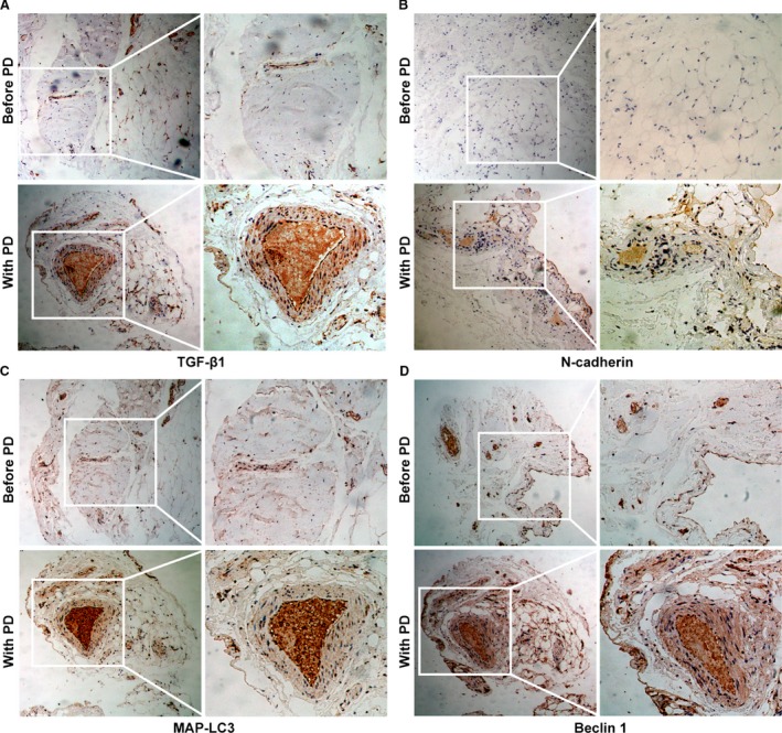
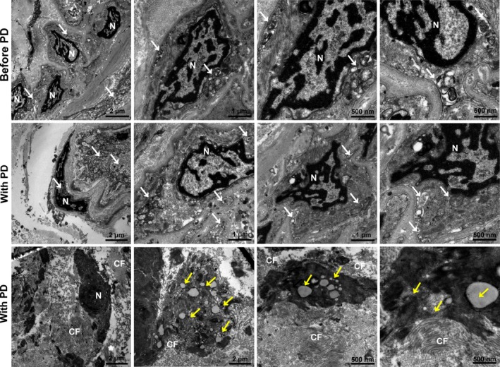
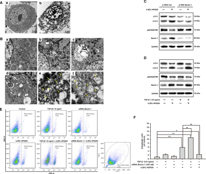
Long-term peritoneal dialysis is accompanied by functional and histopathological alterations in the peritoneal membrane. In the long process of peritoneal dialysis, high-glucose peritoneal dialysis solution (HGPDS) will aggravate the peritoneal fibrosis, leading to decreased effectiveness of peritoneal dialysis and ultrafiltration failure. In this study, we found that the coincidence of elevated TGF-β1 expression, autophagy, apoptosis and fibrosis in peritoneal membrane from patients with peritoneal dialysis. The peritoneal membranes from patients were performed with immunocytochemistry and transmission electron microscopy. Human peritoneal mesothelial cells were treated with 1.5%, 2.5% and 4.25% HGPDS for 24 hrs; Human peritoneal mesothelial cells pre-treated with TGF-β1 (10 ng/ml) or transfected with siRNA Beclin1 were treated with 4.25% HGPDS or vehicle for 24 hrs. We further detected the production of TGF-β1, activation of TGF-β1/Smad2/3 signalling, induction of autophagy, EMT, fibrosis and apoptosis. We also explored whether autophagy inhibition by siRNA targeting Beclin 1 reduces EMT, fibrosis and apoptosis in human peritoneal mesothelial cells. HGPDS increased TGF-β1 production, activated TGF-β1/Smad2/3 signalling and induced autophagy, fibrosis and apoptosis hallmarks in human peritoneal mesothelial cells; HGPDS-induced Beclin 1-dependent autophagy in human peritoneal mesothelial cells; Autophagy inhibition by siRNA Beclin 1 reduced EMT, fibrosis and apoptosis in human peritoneal mesothelial cells. Taken all together, these studies are expected to open a new avenue in the understanding of peritoneal fibrosis, which may guide us to explore the compounds targeting autophagy and achieve the therapeutic improvement of PD.
Keywords: Beclin 1-dependent autophagy; apoptosis; fibrosis; high-glucose peritoneal dialysis solution; human peritoneal mesothelial cells.
© 2017 The Authors. Journal of Cellular and Molecular Medicine published by John Wiley & Sons Ltd and Foundation for Cellular and Molecular Medicine.
Figures


References
-
- Krediet RT, Lindholm B, Rippe B. Pathophysiology of peritoneal membrane failure. Perit Dial Int. 2000; 20: S22–42. - PubMed
-
- Yanez‐Mo M, Lara‐Pezzi E, Selgas R, et al Peritoneal dialysis and epithelial‐to‐mesenchymal transition of mesothelial cells. N Engl J Med. 2003; 348: 403–13. - PubMed
-
- Witowski J, Wisniewska J, Korybalska K, et al Prolonged exposure to glucose degradation products impairs viability and function of human peritoneal mesothelial cells. J Am Soc Nephrol. 2001; 12: 2434–41. - PubMed
Publication types
MeSH terms
Substances
LinkOut - more resources
Full Text Sources
Other Literature Sources
Miscellaneous
