

A medical imaging analysis system for trigger finger using an adaptive texture-based active shape model (ATASM) in ultrasound images
- PMID: 29077737
- PMCID: PMC5659776
- DOI: 10.1371/journal.pone.0187042
A medical imaging analysis system for trigger finger using an adaptive texture-based active shape model (ATASM) in ultrasound images
Abstract
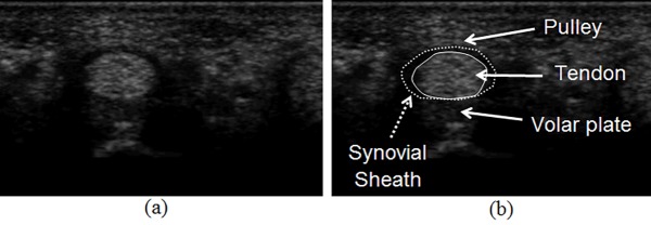
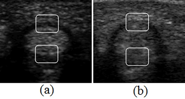
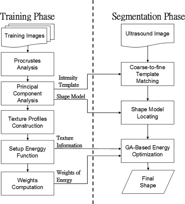
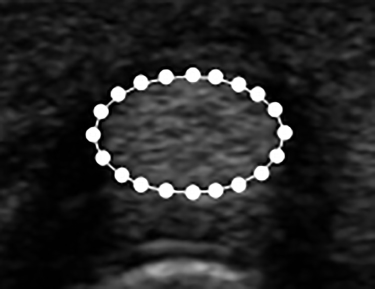
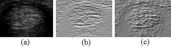
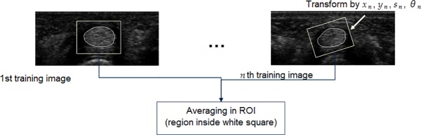
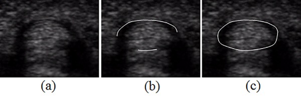
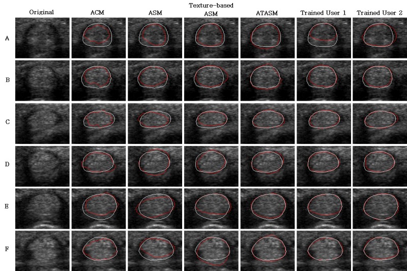
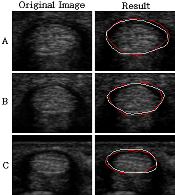
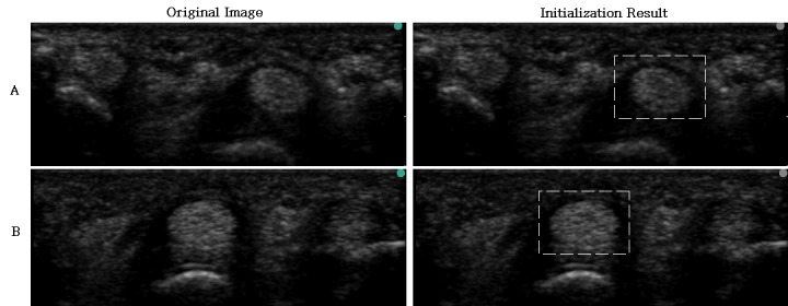
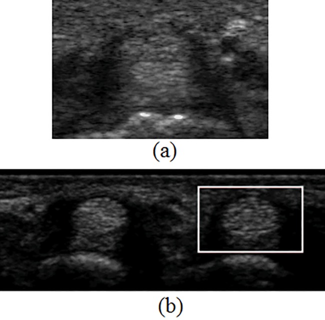
Trigger finger has become a prevalent disease that greatly affects occupational activity and daily life. Ultrasound imaging is commonly used for the clinical diagnosis of trigger finger severity. Due to image property variations, traditional methods cannot effectively segment the finger joint's tendon structure. In this study, an adaptive texture-based active shape model method is used for segmenting the tendon and synovial sheath. Adapted weights are applied in the segmentation process to adjust the contribution of energy terms depending on image characteristics at different positions. The pathology is then determined according to the wavelet and co-occurrence texture features of the segmented tendon area. In the experiments, the segmentation results have fewer errors, with respect to the ground truth, than contours drawn by regular users. The mean values of the absolute segmentation difference of the tendon and synovial sheath are 3.14 and 4.54 pixels, respectively. The average accuracy of pathological determination is 87.14%. The segmentation results are all acceptable in data of both clear and fuzzy boundary cases in 74 images. And the symptom classifications of 42 cases are also a good reference for diagnosis according to the expert clinicians' opinions.
Conflict of interest statement
Figures
References
-
- Ryzewicz M, Wolf JM. Trigger digits: principles, management, and complications. The Journal of hand surgery. 2006. January 31;31(1):135–46. doi: 10.1016/j.jhsa.2005.10.013 - DOI - PubMed
-
- Guerini H, Pessis E, Theumann N, Le Quintrec JS, Campagna R, Chevrot A, et al. Sonographic appearance of trigger fingers. Journal of Ultrasound in Medicine. 2008. October 1;27(10):1407–13. - PubMed
-
- Miyamoto H, Miura T, Isayama H, Masuzaki R, Koike K, Ohe T. Stiffness of the first annular pulley in normal and trigger fingers. The Journal of hand surgery. 2011. September 30;36(9):1486–91. doi: 10.1016/j.jhsa.2011.05.038 - DOI - PubMed
-
- Sato J, Ishii Y, Noguchi H, Takeda M. Sonographic appearance of the flexor tendon, volar plate, and A1 pulley with respect to the severity of trigger finger. The Journal of hand surgery. 2012. October 31;37(10). - PubMed
-
- Gabor D. Theory of communication. Part 1: The analysis of information. Journal of the Institution of Electrical Engineers-Part III: Radio and Communication Engineering. 1946. November 1;93(26):429–41.
MeSH terms
LinkOut - more resources
Full Text Sources
Other Literature Sources
