Review
. 2018 Jan:152:1-14.
doi: 10.1016/j.biomaterials.2017.10.028. Epub 2017 Oct 20.

Bioengineering a non-genotoxic vector for genetic modification of mesenchymal stem cells

Affiliations
Review

Bioengineering a non-genotoxic vector for genetic modification of mesenchymal stem cells

Xuguang Chen et al. Biomaterials. 2018 Jan.

Abstract

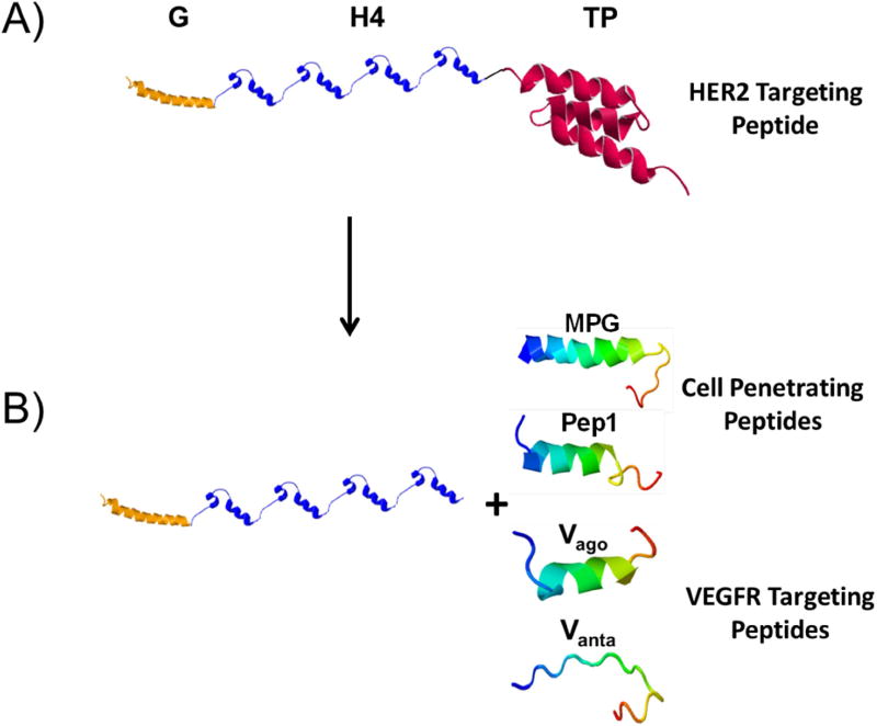
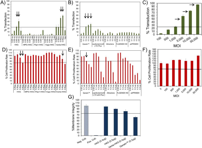
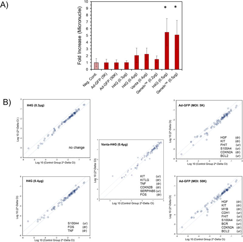
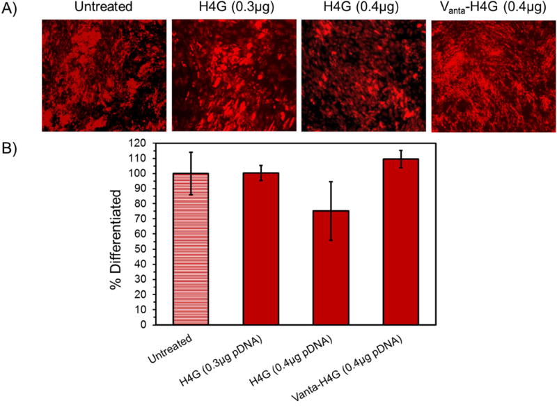
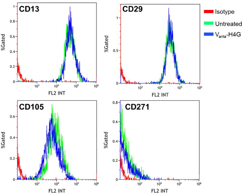
Vectors used for stem cell transfection must be non-genotoxic, in addition to possessing high efficiency, because they could potentially transform normal stem cells into cancer-initiating cells. The objective of this research was to bioengineer an efficient vector that can be used for genetic modification of stem cells without any negative somatic or genetic impact. Two types of multifunctional vectors, namely targeted and non-targeted were genetically engineered and purified from E. coli. The targeted vectors were designed to enter stem cells via overexpressed receptors. The non-targeted vectors were equipped with MPG and Pep1 cell penetrating peptides. A series of commercial synthetic non-viral vectors and an adenoviral vector were used as controls. All vectors were evaluated for their efficiency and impact on metabolic activity, cell membrane integrity, chromosomal aberrations (micronuclei formation), gene dysregulation, and differentiation ability of stem cells. The results of this study showed that the bioengineered vector utilizing VEGFR-1 receptors for cellular entry could transfect mesenchymal stem cells with high efficiency without inducing genotoxicity, negative impact on gene function, or ability to differentiate. Overall, the vectors that utilized receptors as ports for cellular entry (viral and non-viral) showed considerably better somato- and genosafety profiles in comparison to those that entered through electrostatic interaction with cellular membrane. The genetically engineered vector in this study demonstrated that it can be safely and efficiently used to genetically modify stem cells with potential applications in tissue engineering and cancer therapy.

Keywords: Cell transfection; Genotoxicity; Nanoparticles; Non-viral; Stem cells; Vector engineering.


Conflict of interest statement

Conflicts of Interest

None

Figures

Figure 1
Figure 1
The gating protocol that was designed for quantification of micronuclei formation in transfected stem cells.
Figure 2
Figure 2
A) Schematics of the fusion vector composed of a fusogenic peptide GALA (G) to disrupt endosomal membranes, a DNA condensing motif with inherent nuclear localization signal (H4) and a HER2 targeting peptide (TP). B) By removing the HER2 targeting peptide and replacing it with VEGFR targeting or cell penetrating peptides, the vector is tailor-made for carrying genes into MSCs. The 3-D structure of each motif was simulated independently by I-TASSER server for protein structure and function prediction [33].
Figure 3
Figure 3
Characterization of nanoparticles in terms of size, charge, and shape. A) Size of DBV/pEGFP nanocomplexes as determined by dynamic light scattering. B) Surface charge of DBV/pEGFP nanocomplexes as determined by laser Doppler velocimetry. C) Shape of DBV/pEGFP nanocomplexes captured by TEM. The scale bar is 100nm (magnification: 75000×). D) Surface charge analysis of commercial vectors in complex with pEGFP.
Figure 4
Figure 4
Characterization of ADSCs in terms of cell cycle and expression of VEGFR-1. A) Flow cytometry histograms showing the percentage of cells in each phase at different time points (i.e., 16–28 h). B) Bar chart summarizing the percentage of cell population in each cell cycle phase at different time points. As the percentages of cells in Sub G1 phase are very low, they are not observable in the bar chart. C) Flow cytometry histogram/dotplot showing the overexpression of VEGFR-1 on the surface of ADSCs (left panel), A431 cells (middle panel) and in comparison (right panel).
Figure 5
Figure 5
Evaluation of transfection efficiency and impact on cell proliferation rate of DBVs and commercial vectors. A-C) Bar charts that quantitatively demonstrate the percentage of transfected cells using DBVs and commercial non-viral and viral vectors. The arrows point at the most efficient vectors. D-F) Bar charts that demonstrate the impact of DBVs and commercial vectors on the proliferation rate of ADSCs. The arrows highlight the vectors that had high efficiencies (>25%) with acceptable impacts on cell proliferation rate. G) LDH release assay demonstrating the impact of vectors on cell membrane integrity.
Figure 6
Figure 6
A) Evaluation of the impact of vectors on the formation of micronuclei in transfected ADSCs. The percentage of micronuclei in untransfected cells is normalized to a one-fold increase and is considered as the negative control. B) PCR microarray analysis of the dysregulated genes in cells transfected with H4G (0.3 and 0.4 µg pEGFP), Vanta-H4G (0.4 µg pEGFP) and Ad-GFP (MOI: 5K and 50K). Only the upregulated (ur) and downregulated (dr) genes are mentioned in each panel.
Figure 7
Figure 7
ADSC differentiation into adipocyte. A) Fluorescent microscopy images of the differentiated ADSCs. B) Bar chart showing the percentages of differentiated cells in each treated and untreated group using flow cytometry.
Figure 8
Figure 8
Expression of surface markers CD13, CD29, CD105 and CD271 before and after transfection of ADSCs with Vanta-H4G (0.4 µg).

References

    1. Shah K. Mesenchymal stem cells engineered for cancer therapy. Adv Drug Deliv Rev. 2012;64(8):739–48. - PMC - PubMed
    1. Hamada H, Kobune M, Nakamura K, Kawano Y, Kato K, Honmou O, Houkin K, Matsunaga T, Niitsu Y. Mesenchymal stem cells (MSC) as therapeutic cytoreagents for gene therapy. Cancer Sci. 2005;96(3):149–56. - PMC - PubMed
    1. Uhl M, Weiler M, Wick W, Jacobs AH, Weller M, Herrlinger U. Migratory neural stem cells for improved thymidine kinase-based gene therapy of malignant gliomas. Biochem Biophys Res Commun. 2005;328(1):125–9. - PubMed
    1. Kucerova L, Altanerova V, Matuskova M, Tyciakova S, Altaner C. Adipose tissue-derived human mesenchymal stem cells mediated prodrug cancer gene therapy. Cancer Res. 2007;67(13):6304–13. - PubMed
    1. Cavarretta IT, Altanerova V, Matuskova M, Kucerova L, Culig Z, Altaner C. Adipose tissue-derived mesenchymal stem cells expressing prodrug-converting enzyme inhibit human prostate tumor growth. Mol Ther. 2010;18(1):223–31. - PMC - PubMed

Substances