

Intratumor and Intertumor Heterogeneity in Melanoma
- PMID: 29078205
- PMCID: PMC5671412
- DOI: 10.1016/j.tranon.2017.09.007
Intratumor and Intertumor Heterogeneity in Melanoma
Abstract
Melanoma is a cancer that exhibits one of the most aggressive and heterogeneous features. The incidence rate escalates. A high number of clones harboring various mutations contribute to an exceptional level of intratumor heterogeneity of melanoma. It also refers to metastases which may originate from different subclones of primary lesion. Such component of the neoplasm biology is termed intertumor and intratumor heterogeneity. These levels of tumor heterogeneity hinder accurate diagnosis and effective treatment. The increasing number of research on the topic reflects the need for understanding limitation or failure of contemporary therapies. Majority of analyses concentrate on mutations in cancer-related genes. Novel high-throughput techniques reveal even higher degree of variations within a lesion. Consolidation of theories and researches indicates new routes for treatment options such as targets for immunotherapy. The demand for personalized approach in melanoma treatment requires extensive knowledge on intratumor and intertumor heterogeneity on the level of genome, transcriptome/proteome, and epigenome. Thus, achievements in exploration of melanoma variety are described in details. Particularly, the issue of tumor heterogeneity or homogeneity given BRAF mutations is discussed.
Copyright © 2017 The Authors. Published by Elsevier Inc. All rights reserved.
Figures
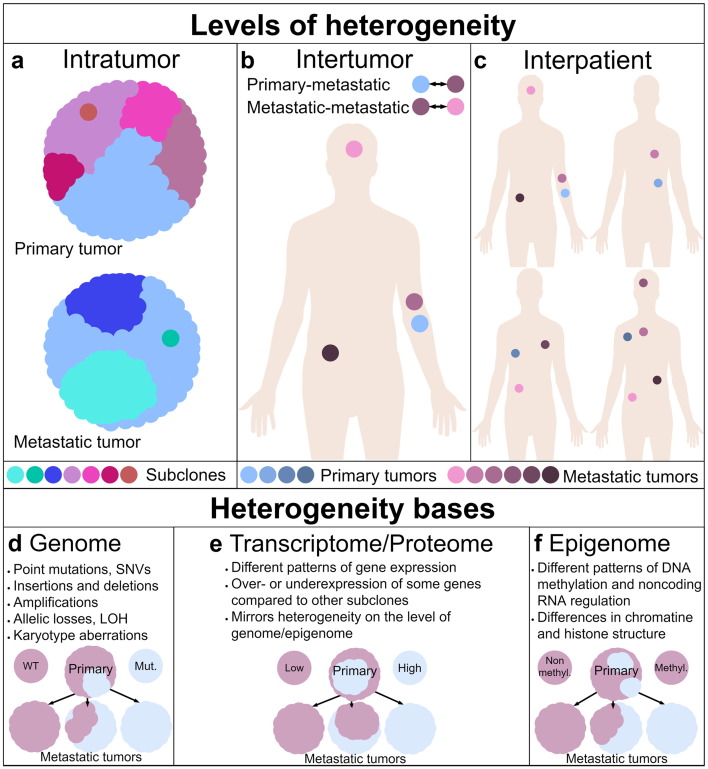
 ) in the primary tumor (top) was also the founder of metastases (bottom). (B) The differences among the primary tumor () and metastatic (
 ) tumors and among metastases constitute intrapatient intertumor heterogeneity. (C) The differences among tumors from different patients are termed interpatient heterogeneity. Heterogeneity bases. (D) Genetic heterogeneity arises from various changes within genome. On this scheme, the primary tumor (top) contains cells wild type given some gene as well as cells harboring a mutated allele. Wild-type subclone was the founder of two metastases (bottom left). However, one of them acquired a mutation in this gene (middle). The mutated subclone was the founder of third metastasis. (E) Heterogeneity of transcriptome and proteome constitute heterogeneity of gene expression. Low-expressing subclone was the founder of one metastasis, which is homogeneous given this gene expression. However, the other two metastatic tumors were formed by a high-expressing subclone, although both metastases heterogeneously express this gene on mRNA/protein marker level. (F) The primary tumor is homogeneous given methylation status of some gene promoter – remain nonmethylated. One metastasis mirrors the status of primary tumor, while the other two metastatic tumors are heterogeneous. One tumor (middle) exhibits epigenetic heterogeneity developed during progression since one of the tumor cells acquired an epimutation. The last metastasis is homogeneous because the founder cell acquired an epimutation. Therefore, methylation of promoter is present in all tumor cells.
References
-
- Dick JE. Stem cell concepts renew cancer research. Blood. 2008;112(13):4793–4807. - PubMed
-
- Nassar A, Radhakrishnan A, Cabrero IA, Cotsonis GA, Cohen C. Intratumoral heterogeneity of immunohistochemical marker expression in breast carcinoma: a tissue microarray-based study. Appl Immunohistochem Mol Morphol. 2010;18(5):433–441. - PubMed
-
- Coons SW, Johnson PC, Shapiro JR. Cytogenetic and flow cytometry DNA analysis of regional heterogeneity in a low grade human glioma. Cancer Res. 1995;55(7):1569–1577. - PubMed
Publication types
LinkOut - more resources
Full Text Sources
Other Literature Sources
Research Materials
