. 2017 Oct 31;114(44):11775-11780.
doi: 10.1073/pnas.1707863114. Epub 2017 Oct 19.

Osteocyte calcium signals encode strain magnitude and loading frequency in vivo

Affiliations

Osteocyte calcium signals encode strain magnitude and loading frequency in vivo

Karl J Lewis et al. Proc Natl Acad Sci U S A. .

Abstract

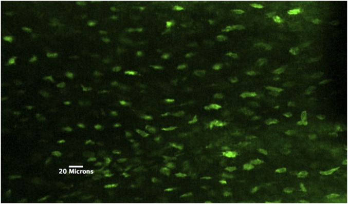
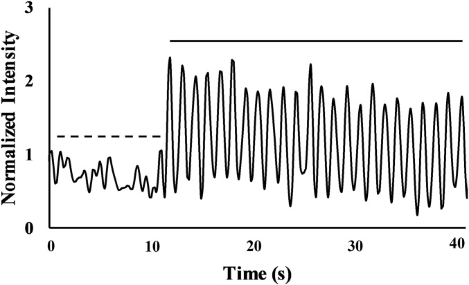
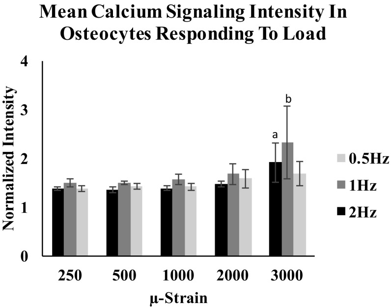
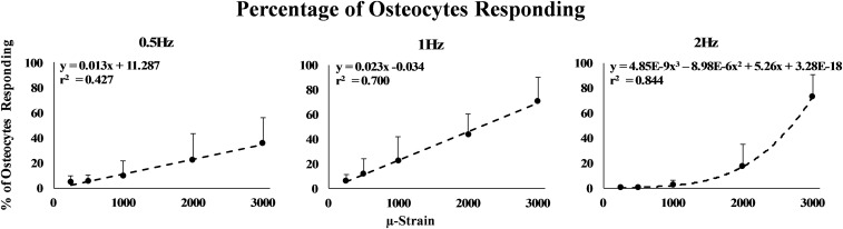
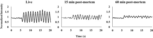
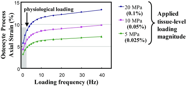
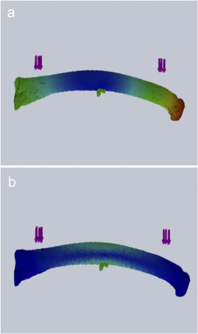
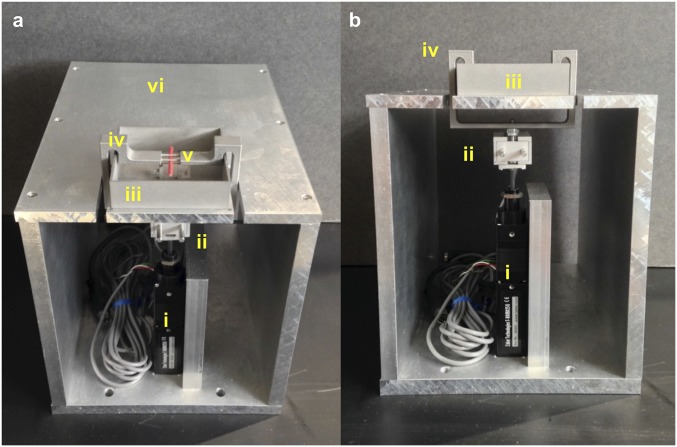
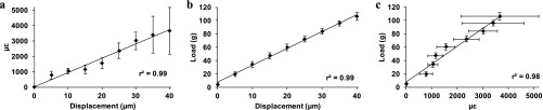
Osteocytes are considered to be the major mechanosensory cells of bone, but how osteocytes in vivo process, perceive, and respond to mechanical loading remains poorly understood. Intracellular calcium (Ca2+) signaling resulting from mechanical stimulation has been widely studied in osteocytes in vitro and in bone explants, but has yet to be examined in vivo. This is achieved herein by using a three-point bending device which is capable of delivering well-defined mechanical loads to metatarsal bones of living mice while simultaneously monitoring the intracellular Ca2+ responses of individual osteocytes by using a genetically encoded fluorescent Ca2+ indicator. Osteocyte responses are imaged by using multiphoton fluorescence microscopy. We investigated the in vivo responses of osteocytes to strains ranging from 250 to 3,000 [Formula: see text] and frequencies from 0.5 to 2 Hz, which are characteristic of physiological conditions reported for bone. At all loading frequencies examined, the number of responding osteocytes increased strongly with applied strain magnitude. However, Ca2+ intensity within responding osteocytes did not change significantly with physiological loading magnitudes. Our studies offer a glimpse into how these critical bone cells respond to mechanical load in vivo, as well as provide a technique to determine how the cells encode magnitude and frequency of loading.

Keywords: bone; calcium signaling; in vivo loading; mechanotransduction; osteocytes.


Conflict of interest statement

The authors declare no conflict of interest.

Figures

Fig. 1.
Fig. 1.
Multiphoton z-stack image of OtGP3 MT3 osteocytes in vivo. Note that all cells in the field of view exhibit GCaMP3 signal, indicating highly efficient expression in osteocytes.
Fig. 2.
Fig. 2.
A representative Ca2+ trace for a single osteocyte before (dashed line) and after the start of cyclical loading (solid line) at 1 Hz to 3,000 μϵ. Before mechanical loading, all osteocytes exhibited low-level Ca2+ fluctuations. Immediately following the start of mechanical loading, responsive cells showed cyclic increases in Ca2+ fluorescence intensity coinciding with the applied loading frequency.
Fig. 3.
Fig. 3.
Intensity of Ca2+ signaling per osteocytes was not changed with increasing strain levels up to 2,000 μϵ in all loading rate groups. Increases in Ca2+ were noted at 3,000 μϵ. (a) P< 0.05; 2-Hz loading group; (b) P< 0.05 1-Hz loading group. Error bars represent SD, n = 6 per group.
Fig. 4.
Fig. 4.
Shown is the percentage of responding osteocytes as a function of applied strain magnitude for 0.5-, 1-, and 2-Hz loading frequencies. All response curves increased with increasing strain and were effectively linear at 0.5- and 1-Hz loading. The 2-Hz loading response curve was nonlinear. Error bars represent SD. n = 6 per frequency group.
Fig. 5.
Fig. 5.
Typical Ca2+ traces for two representative osteocytes before and after the onset of mechanical loading in live bone (Left) and at 15 min (Center) and 60 min (Right) postmortem. Osteocyte Ca2+ signal amplitude acutely after death was markedly attenuated compared with in vivo, although the Ca2+ signaling still tracked the applied loading frequency. In contrast, at 60 min postmortem, the response of osteocytes to loading was low and irregular, with Ca2+ signaling no longer consistent with the applied loading frequency. n = 3 per group.
Fig. 6.
Fig. 6.
Predicted axial strain at the transduction sites on the osteocyte process cell membrane as a function of loading frequency at various tissue-loading magnitudes [adapted from the mathematical model of Wang et al. (5)]. The shaded region has been added to highlight the exponential increases in focal membrane strains expected on osteocyte process over the physiological frequency range examined experimentally in the present studies.
Fig. S1.
Fig. S1.
FEA modeling results with heat maps of total displacement (A) and von Mises strain (B). Note that the location of maximum strain coincides with the area showing little z axis displacement.
Fig. S2.
Fig. S2.
Angled (A) and frontal (B) views of custom device for in vivo loading studies. Actuator (i) and load cell (ii) are in series beneath the platform and rectangular water bath (iii) to contain the foot. Upper loading contacts for three-point bending are part of the arm bracket assembly (iv) that wraps around the water bath, and is pulled from below to apply loads to the dorsal bone surface. The red stick shown in the apparatus represents the positioning of the MT3 in the device (v). Mice are under anesthesia while on the platform (vi). The entire apparatus sits on the stage of a multiphoton microscope.
Fig. S3.
Fig. S3.
Calibration curves from MT3 bones showing strain vs. displacement (A), load vs. displacement (B), and load vs. strain (C). Error bars represent SD, n = 6.

Similar articles

Cited by

References

    1. Cabahug-Zuckerman P, et al. Osteocyte apoptosis caused by Hindlimb unloading is required to trigger osteocyte RANKL production and subsequent resorption of cortical and Trabecular bone in mice femurs. J Bone Miner Res. 2016;31:1356–1365. - PMC - PubMed
    1. Plotkin LI, et al. Inhibition of osteocyte apoptosis prevents the increase in osteocytic receptor activator of nuclear factor κB ligand (RANKL) but does not stop bone resorption or the loss of bone induced by unloading. J Biol Chem. 2015;290:18934–18942. - PMC - PubMed
    1. Emerton KB, et al. Osteocyte apoptosis and control of bone resorption following ovariectomy in mice. Bone. 2010;46:577–583. - PMC - PubMed
    1. Kennedy OD, et al. Activation of resorption in fatigue-loaded bone involves both apoptosis and active pro-osteoclastogenic signaling by distinct osteocyte populations. Bone. 2012;50:1115–1122. - PMC - PubMed
    1. Wang Y, McNamara LM, Schaffler MB, Weinbaum S. A model for the role of integrins in flow induced mechanotransduction in osteocytes. Proc Natl Acad Sci USA. 2007;104:15941–15946. - PMC - PubMed

Publication types