. 2017 Nov 7;114(45):E9665-E9674.
doi: 10.1073/pnas.1708568114. Epub 2017 Oct 23.

Lack of BACE1 S-palmitoylation reduces amyloid burden and mitigates memory deficits in transgenic mouse models of Alzheimer's disease

Affiliations

Lack of BACE1 S-palmitoylation reduces amyloid burden and mitigates memory deficits in transgenic mouse models of Alzheimer's disease

Robert J Andrew et al. Proc Natl Acad Sci U S A. .

Abstract

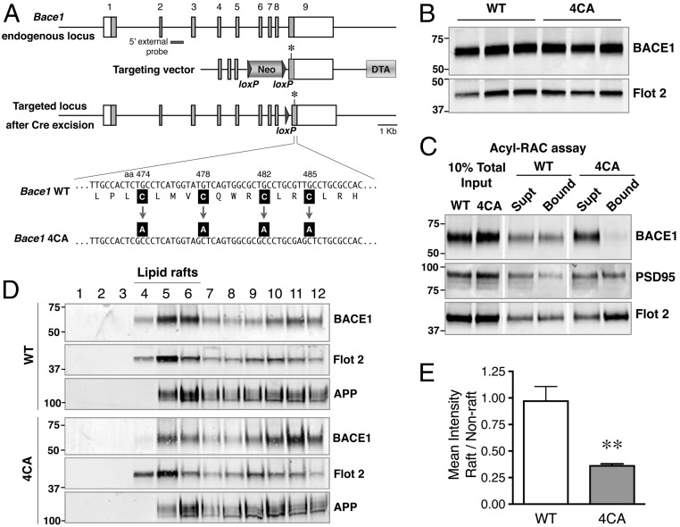
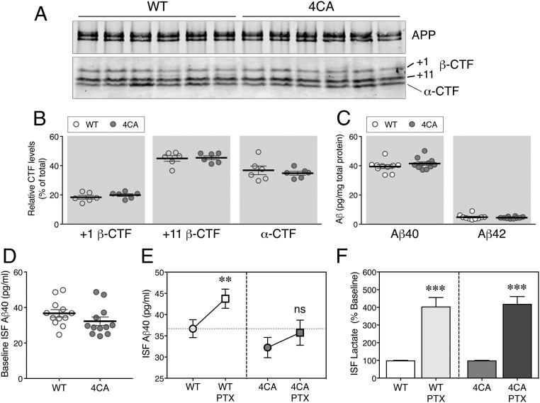
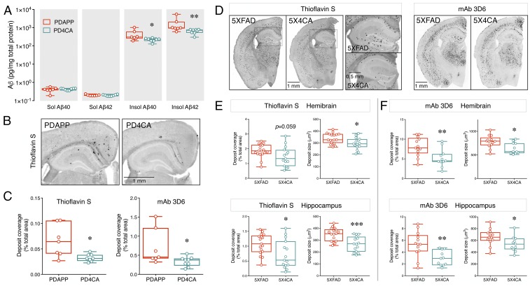
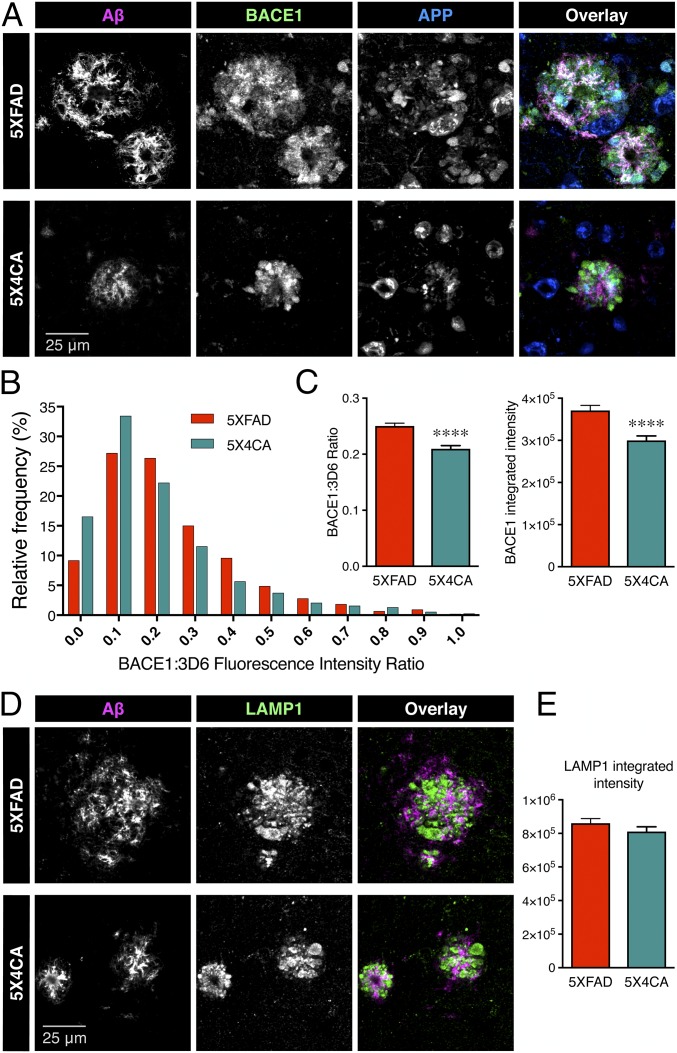
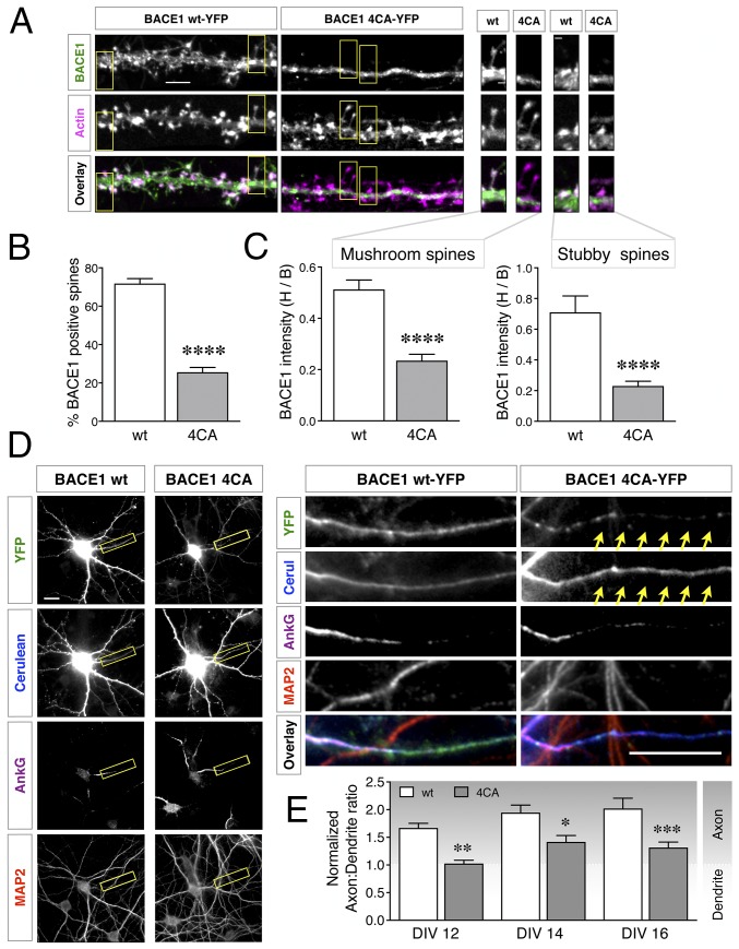
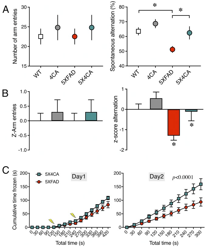
Alzheimer's disease (AD) is a devastating neurodegenerative disorder characterized by pathological brain lesions and a decline in cognitive function. β-Amyloid peptides (Aβ), derived from proteolytic processing of amyloid precursor protein (APP), play a central role in AD pathogenesis. β-Site APP cleaving enzyme 1 (BACE1), the transmembrane aspartyl protease which initiates Aβ production, is axonally transported in neurons and accumulates in dystrophic neurites near cerebral amyloid deposits in AD. BACE1 is modified by S-palmitoylation at four juxtamembrane cysteine residues. S-palmitoylation is a dynamic posttranslational modification that is important for trafficking and function of several synaptic proteins. Here, we investigated the in vivo significance of BACE1 S-palmitoylation through the analysis of knock-in mice with cysteine-to-alanine substitution at the palmitoylated residues (4CA mice). BACE1 expression, as well as processing of APP and other neuronal substrates, was unaltered in 4CA mice despite the lack of BACE1 S-palmitoylation and reduced lipid raft association. Whereas steady-state Aβ levels were similar, synaptic activity-induced endogenous Aβ production was not observed in 4CA mice. Furthermore, we report a significant reduction of cerebral amyloid burden and BACE1 accumulation in dystrophic neurites in the absence of BACE1 S-palmitoylation in mouse models of AD amyloidosis. Studies in cultured neurons suggest that S-palmitoylation is required for dendritic spine localization and axonal targeting of BACE1. Finally, the lack of BACE1 S-palmitoylation mitigates cognitive deficits in 5XFAD mice. Using transgenic mouse models, these results demonstrate that intrinsic posttranslational S-palmitoylation of BACE1 has a significant impact on amyloid pathogenesis and the consequent cognitive decline.

Keywords: 5XFAD; PDAPP; axonal transport; dystrophic neurite; neurodegeneration.


Conflict of interest statement

Conflict of interest statement: D.M.H. is cofounder of C2N Diagnostics, LLC, serves on the scientific advisory board of C2N Diagnostics, and consults for Eli Lilly, AbbVie, GlaxoSmithKline, Genentech, Proclara Biosciences, and Denali.

Figures

Fig. 1.
Fig. 1.
Generation of 4CA knock-in mice. (A, Top) Schematic representation of the Bace1 locus showing the nine exons (numbered); coding regions are shown as filled gray boxes. (Middle) The homology arms of the targeting vector are shown along with the loxP-flanked PGK-Neo and the Diptheria Toxin A (DTA) selection cassettes. An asterisk indicates a silent mutation introduced to generate a unique SacI site in the mutant allele. (Bottom) Schematic structure of the targeted allele after Cre-mediated excision of the PGK-Neo gene. The cysteine-to-alanine substitutions are indicated. (B) Total brain homogenates of WT and 4CA mice (P7) were analyzed by immunoblotting. (C) Detection of S-palmitoylated BACE1 in mouse brain by acyl-RAC. Mouse brain lysates (P7) were subjected to acyl-RAC, and aliquots of captured proteins (Bound) and unbound fraction (Supt) were analyzed by immunoblotting for BACE1, PSD95, and Flotillin 2. (D) P7 mouse brain was homogenized in a buffer containing 0.5% Lubrol WX at 4 °C for 30 min. The lysates were then subject to flotation sucrose density gradient, and an equal volume of each fraction, harvested from the top, was analyzed by immunoblotting. Fractions 4 to 6 represent the interface between 5% and 35% sucrose in the gradient and are enriched in lipid raft marker Flotillin-2. (E) Quantification of the relative distribution of BACE1 in lipid raft and nonraft fractions. **P < 0.01.
Fig. 2.
Fig. 2.
The increase of ISF Aβ in response to neuronal activity is diminished in 4CA mice. (A) Full-length APP and APP CTFs in 2-mo-old WT and 4CA mouse forebrain homogenates were immunoprecipitated, subjected to dephosphorylation, and analyzed by immunoblotting. (B) The signal intensities of +1 or +11 β-CTFs and α-CTFs normalized to total APP CTFs were quantified (n = 6 WT and 6 4CA). (C) The steady-state levels of Aβ40 and Aβ42 in SDS homogenates of forebrains from 2-mo-old animals were analyzed using a V-PLEX 4G8 immunoassay (n = 10 WT and 11 4CA). (D) ISF samples were collected from 7- to 8-mo-old WT and 4CA mice (n = 12 each) by in vivo microdialysis over 15 h and analyzed by ELISA to quantify the steady-state ISF Aβ40 levels. (E) After establishing the baseline, 25 µM PTX was administered by reverse microdialysis to stimulate neuronal activity. The baseline and PTX treatment-induced Aβ40 levels were quantified and analyzed by two-way repeated measure ANOVA. ns, nonsignificant (n = 12 each). (F) The PTX-induced increase in the levels of ISF lactate, an indicator of neuronal activity, was quantified from the same samples as above. **P < 0.01; ***P < 0.001.
Fig. 3.
Fig. 3.
Reduced amyloid deposition in AD mouse models in the absence of BACE1 S-palmitoylation. (A) Forebrain tissue harvested from 9-mo-old female PDAPP and PD4CA mice (n = 7 and 8, respectively) were sequentially homogenized in PBS and guanidine to extract soluble (Sol) and insoluble (Insol) Aβ. The levels of Aβ40 and Aβ42 in each fraction were quantified by ELISA. (B) Representative images of fibrillary Aβ deposit staining using Thioflavin S. (C) Quantification of amyloid load in hemibrain by Thioflavin S staining (n = 7 PDAPP; 8 PD4CA) and mAb 3D6 immunostaining (n = 7 PDAPP; 9 PD4CA). (D) Representative images of 4-mo-old 5XFAD and 5X4CA mice analyzed by Thioflavin S staining and mAb 3D6 immunostaining. A higher magnification of the boxed region is shown. (E and F) Quantification of amyloid load and deposit size in hemibrain and hippocampus by Thioflavin S staining (n = 17 5XFAD; 15 5X4CA) and mAb 3D6 immunostaining (n = 13 5XFAD; 11 5X4CA). *P < 0.05; **P < 0.01; ***P < 0.001.
Fig. 4.
Fig. 4.
Reduced accumulation of BACE1 in dystrophic neurites in the absence of BACE1 S-palmitoylation. (A) Representative images of immunofluorescence labeling of Aβ deposits (mAb 3D6, magenta), APP (blue), and BACE1 (green). (B) Frequency distribution analysis of BACE1 fluorescence intensity relative to that of Aβ in individual amyloid deposits. All nonoverlapping deposits were counted within equal areas of four cortical regions and the hippocampus (n = 5XFAD 1,782 deposits from four mice; 5X4CA 1,443 deposits from four mice). (C) The graphs represent the mean ratio of BACE1 to Aβ fluorescence intensity and the mean integrated intensity of BACE1 immunostaining. (D) Representative images of immunofluorescence labeling of Aβ deposits (mAb 3D6, magenta) and LAMP1 (green). (E) Integrated intensity of LAMP1 staining in nonoverlapping deposits in four cortical regions and the hippocampus (n = 5XFAD 1,417 deposits from four mice; 5X4CA 915 deposits from four mice). ****P < 0.0001.
Fig. 5.
Fig. 5.
Modulation of BACE1 trafficking in neurons by S-palmitoylation. (A) Hippocampal neurons were cotransfected with mApple-Actin and either YFP-tagged BACE1 wt or 4CA constructs. Neurons were fixed 4 d later (DIV15) and analyzed by microscopy. A higher magnification of the boxed regions depicting mushroom or stubby spines are shown on the Right. (Scale bars: Left, 5 µm; Right, 1 µm.) (B) Quantification of the percentage of actin-positive spines that contain BACE1 (n = 23 WT and 24 4CA neurons from three independent cultures). (C) Quantification of BACE1 intensity in spine head relative to the base of the spine (H/B) (n = WT 279 and 4CA 300 mushroom spines; WT 61 and 4CA 67 stubby spines). (D) Neurons coexpressing Cerulean and BACE1-YFP were fixed at DIV16 and immunostained for AnkG and MAP2, to identify the axon initial segment and dendrites, respectively. A higher magnification of the boxed region is shown on the Right. The arrows highlight the axon. (Scale bars, 20 μm.) (E) Neurons were transfected on DIV10, and nADRs were calculated on DIV12 (n = 30 neurons each), DIV14 (n = 30 neurons each), or DIV16 (n = WT 34 and 4CA 35 neurons). Results are from at least three independent cultures, each analyzed in parallel on all time points. *P < 0.05; **P < 0.01; ***P < 0.001; ****P < 0.0001.
Fig. 6.
Fig. 6.
The lack of BACE1 S-palmitoylation mitigates cognitive deficits in 5XFAD mice. (A) Cohorts of mice of the indicated genotypes were assessed for spatial working memory in the Y-maze test. The number of arm entries and the spontaneous alternation behavior were quantified (n = WT 14; 4CA 12; 5XFAD 15; 5X4CA 11 mice). (B) The normalized z-scores of the test results were calculated to assess the significance of the differences observed in the different cohorts of mice. (C) Hippocampal-dependent associative learning and memory task. 5XFAD and 5X4CA animals were exposed to the contextual fear conditioning paradigm, and freezing behavior was measured either during the training (day 1) or 24 h after the conditioning (day 2) (n = 5XFAD 19; 5X4CA 12 mice). The lightning bolts indicate the time when the electric shocks were delivered. *P < 0.05.

References

    1. Thinakaran G, Koo EH. Amyloid precursor protein trafficking, processing, and function. J Biol Chem. 2008;283:29615–29619. - PMC - PubMed
    1. Mullan M, et al. A pathogenic mutation for probable Alzheimer’s disease in the APP gene at the N-terminus of beta-amyloid. Nat Genet. 1992;1:345–347. - PubMed
    1. Citron M, et al. Mutation of the beta-amyloid precursor protein in familial Alzheimer’s disease increases beta-protein production. Nature. 1992;360:672–674. - PubMed
    1. Cai XD, Golde TE, Younkin SG. Release of excess amyloid beta protein from a mutant amyloid beta protein precursor. Science. 1993;259:514–516. - PubMed
    1. Di Fede G, et al. A recessive mutation in the APP gene with dominant-negative effect on amyloidogenesis. Science. 2009;323:1473–1477. - PMC - PubMed

Publication types

MeSH terms

LinkOut - more resources