. 2017 Nov 7;114(45):E9512-E9519.
doi: 10.1073/pnas.1710702114. Epub 2017 Oct 25.

Bacteriorhodopsin-like channelrhodopsins: Alternative mechanism for control of cation conductance

Affiliations

Bacteriorhodopsin-like channelrhodopsins: Alternative mechanism for control of cation conductance

Oleg A Sineshchekov et al. Proc Natl Acad Sci U S A. .

Abstract

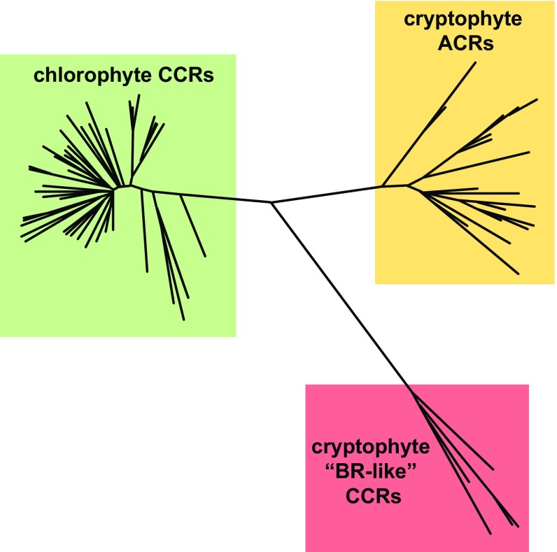
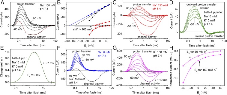
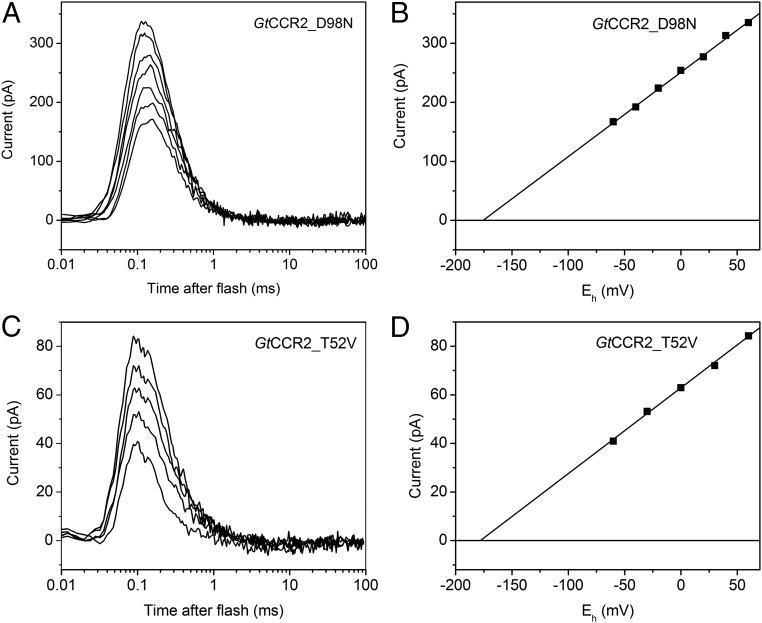
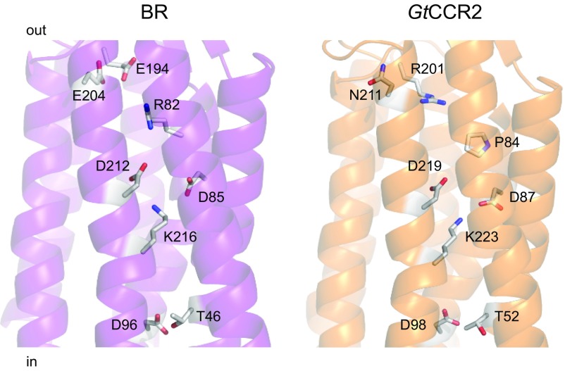
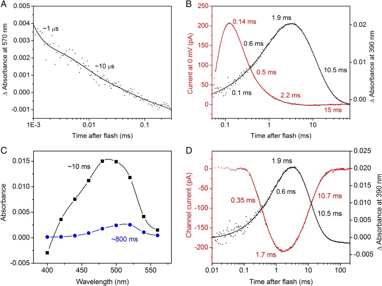
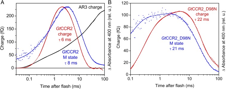
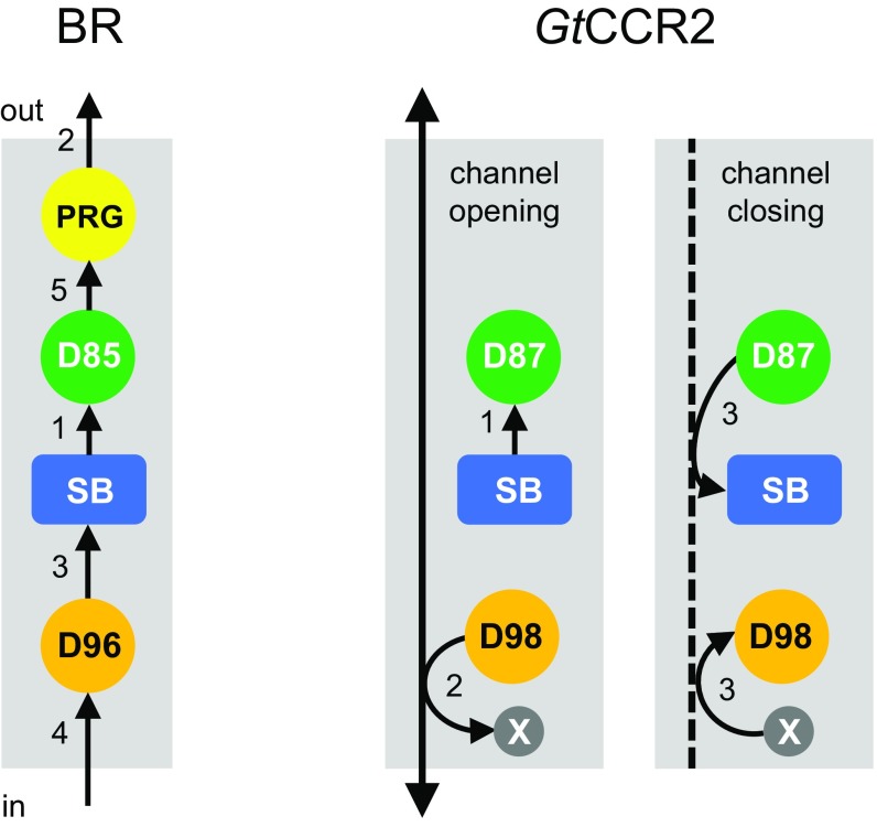
The recently discovered cation-conducting channelrhodopsins in cryptophyte algae are far more homologous to haloarchaeal rhodopsins, in particular the proton pump bacteriorhodopsin (BR), than to earlier known channelrhodopsins. They uniquely retain the two carboxylate residues that define the vectorial proton path in BR in which Asp-85 and Asp-96 serve as acceptor and donor, respectively, of the photoactive site Schiff base (SB) proton. Here we analyze laser flash-induced photocurrents and photochemical conversions in Guillardia theta cation channelrhodopsin 2 (GtCCR2) and its mutants. Our results reveal a model in which the GtCCR2 retinylidene SB chromophore rapidly deprotonates to the Asp-85 homolog, as in BR. Opening of the cytoplasmic channel to cations in GtCCR2 requires the Asp-96 homolog to be unprotonated, as has been proposed for the BR cytoplasmic channel for protons. However, reprotonation of the GtCCR2 SB occurs not from the Asp-96 homolog, but by proton return from the earlier protonated acceptor, preventing vectorial proton translocation across the membrane. In GtCCR2, deprotonation of the Asp-96 homolog is required for cation channel opening and occurs >10-fold faster than reprotonation of the SB, which temporally correlates with channel closing. Hence in GtCCR2, cation channel gating is tightly coupled to intramolecular proton transfers involving the same residues that define the vectorial proton path in BR.

Keywords: channelrhodopsins; ion transport; optogenetics; photocycle; proton transfers.


Conflict of interest statement

The authors declare no conflict of interest.

Figures

Fig. 1.
Fig. 1.
A phylogenetic tree of three families of channelrhodopsins constructed by the neighbor-joining method. Approximately ∼60 chlorophyte CCRs and ∼30 cryptophyte АCRs have been identified to date. Only 5 cryptophyte CCRs have been characterized by heterologous expression, but the fully sequenced G. theta genome harbors several other homologous genes, and more homologs are likely to be found in other cryptophyte species.
Fig. 2.
Fig. 2.
Two components of GtCCR2 photocurrents under single-turnover conditions and their ionic dependencies. (A, C, D, F, and G) Representative series of current traces recorded from wild-type GtCCR2 in response to a 6-ns laser flash at different ionic conditions. The main ions in the bath solution are indicated in the panels (for other components see Materials and Methods); the pipette solution was standard except in D and E, in which it contained no Na+, K+, or Ca2+. The holding voltage at the amplifier output was changed in 20-mV (A, C, F, and G) or 30-mV (D) steps from −60 mV. In A and G, multiexponential fits of experimental data recorded at −60 mV are shown as the dashed lines. In F, photocurrent recorded from the same cell at −60 mV with 150 mM Na+ in the bath is shown for comparison as the black line. (B) The voltage dependence of the fast (squares) and channel currents corrected for inward proton transfer (circles) at pH 7.4 (black) and 5.4 (red) of the bath medium in the presence of 150 mM Na+. For 0 mM Na+ (blue), only the scaled values of the fast current are shown, because no channel currents were detected under these conditions. The straight lines show linear fits to the data. The arrow shows a shift in Erev measured at 150 mM Na+ upon acidification of the medium from pH 7.4 to 5.4. (E) Kinetics of charge transfer calculated as the area under the current curve measured in the absence of any channel activity. (H) The voltage dependence of the channel current at 50 (empty symbols) or 150 (filled symbols) mM K+ in the bath, corrected for inward proton transfer. To calculate the error values, data for E and H obtained on different cells were normalized and plotted as relative units (rel. u.).
Fig. 3.
Fig. 3.
Role of the homologs of the proton donor in BR and its hydrogen-bonded threonine in channel activity of GtCCR2. (A and C) Series of current traces recorded in response to a 6-ns laser flash at standard conditions from GtCCR2_D98N (A) and GtCCR2_T52V (C). No channel current was detected. (B and D) Corresponding voltage dependences of the peak amplitudes of proton transfer currents.
Fig. 4.
Fig. 4.
A crystal structure of BR (1c3w; Left) and a homology model of GtCCR2 built by the Robetta server using a structure of a haloarchaeal sensory rhodopsin II (2ksy) as a template (Right). The side chains of the key residues involved in intramolecular proton transfers are shown as sticks.
Fig. 5.
Fig. 5.
Laser flash-induced absorbance changes in purified of GtCCR2 and their correlation with photocurrents. (A) Biphasic decay of the K intermediate. The dots show experimental data, and the solid line, a multiexponential fit. (B) The photocurrent trace recorded in the absence of Na+ at 0 mV holding potential is shown as red dots, and kinetics of the M intermediate, as black dots. The results of multiexponential fitting are shown as solid lines; the numbers are the τ-values derived from the fit. (C) The spectral dependence of the amplitudes of the two slowest components derived by global fit. (D) Correlation of the opening and closing of the channel (red) with the appearance and decay of the M intermediate (black). The channel current was calculated by subtraction of the properly scaled proton transfer current (obtained at the positive holding potentials) from the photoelectric signal measured at −60 mV in the standard bath.
Fig. 6.
Fig. 6.
Temporal correlation of the charge transfer and M formation in wild-type GtCCR2 and its D98N mutant. (A) Charge transfer in GtCCR2 (red trace) expressed in HEK293 cells recorded in response to a 6-ns laser flash, and the kinetics of the M intermediate in purified GtCCR2 (blue trace). Charge transfer in the proton pump Arch (black trace) is shown for comparison. (B) Charge transfer (red trace) recorded in response to a 6-ns laser flash and the M intermediate kinetics (blue trace) in the GtCCR2_D98N mutant. Absorption changes in panels are plotted in relative units (rel. u.) to compensate for different expression levels and normalized to the amplitudes of charge transfer plotted as femtocoulombs (fQ).
Fig. 7.
Fig. 7.
Schematic presentation of intramolecular proton transfers in GtCCR2 compared with BR. As reviewed in refs. and , in BR deprotonation of the SB to Asp-85 via a water molecule (1) is followed by a fast release of another proton to the extracellular medium (2). The SB is then reprotonated from Asp-96 whose drop in pKa is facilitated by breakage of its hydrogen bond with Thr-46 (3), and which in turn takes up a proton from the cytoplasm (4). Finally, Asp-85 donates a proton to the proton release group (5). In GtCCR2, the SB is deprotonated to Asp-87 (1), whereas deprotonation of Asp-98 to an unidentified residue or water molecule is coupled to channel opening (2). Return of the proton from Asp-87 to the SB and a presumably synchronous reprotonation of Asp-98 are coupled to channel closing (3). PRG, proton release group; SB, Schiff base; X, an unidentified residue or water molecule.

References

    1. Ernst OP, et al. Microbial and animal rhodopsins: Structures, functions, and molecular mechanisms. Chem Rev. 2014;114:126–163. - PMC - PubMed
    1. Govorunova EG, Sineshchekov OA, Li H, Spudich JL. Microbial rhodopsins: Diversity, mechanisms, and optogenetic applications. Annu Rev Biochem. 2017;86:845–872. - PMC - PubMed
    1. Sineshchekov OA, Jung K-H, Spudich JL. Two rhodopsins mediate phototaxis to low- and high-intensity light in Chlamydomonas reinhardtii. Proc Natl Acad Sci USA. 2002;99:8689–8694. - PMC - PubMed
    1. Nagel G, et al. Channelrhodopsin-1: A light-gated proton channel in green algae. Science. 2002;296:2395–2398. - PubMed
    1. Nagel G, et al. Channelrhodopsin-2, a directly light-gated cation-selective membrane channel. Proc Natl Acad Sci USA. 2003;100:13940–13945. - PMC - PubMed

Publication types

LinkOut - more resources