. 2017 Nov 21;114(47):12396-12400.
doi: 10.1073/pnas.1714896114. Epub 2017 Oct 23.

Abiotic production of sugar phosphates and uridine ribonucleoside in aqueous microdroplets

Affiliations

Abiotic production of sugar phosphates and uridine ribonucleoside in aqueous microdroplets

Inho Nam et al. Proc Natl Acad Sci U S A. .

Abstract

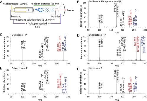
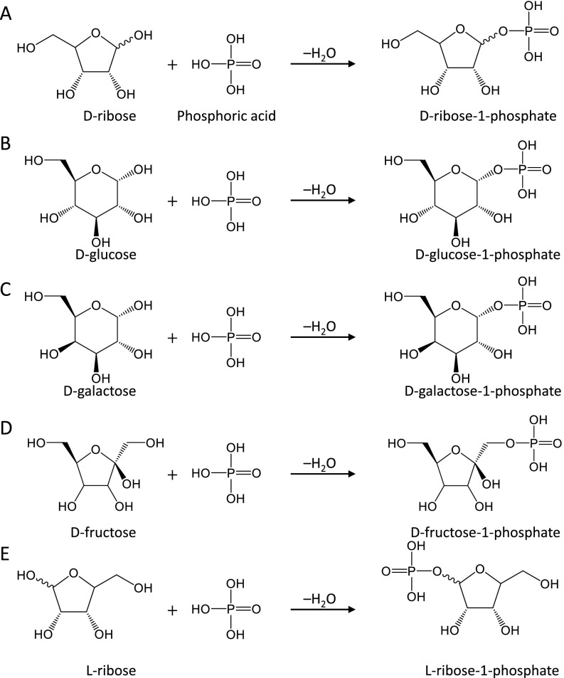
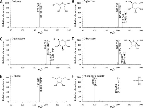
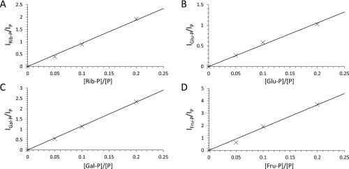
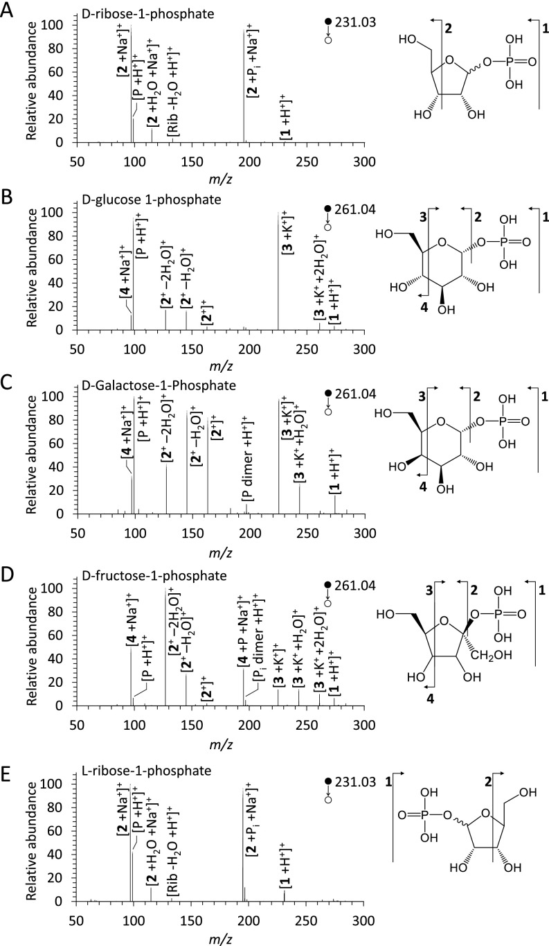
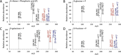
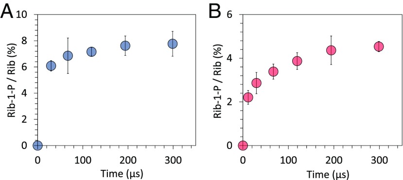
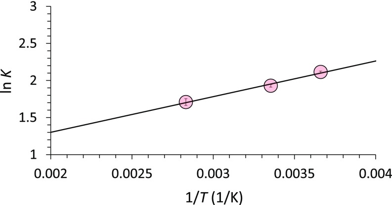
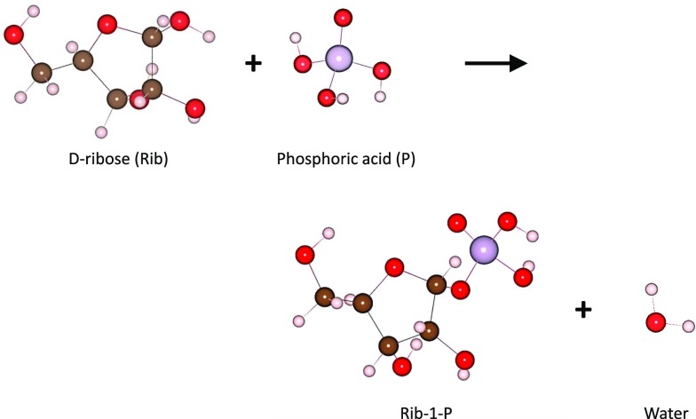
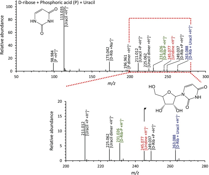
Phosphorylation is an essential chemical reaction for life. This reaction generates fundamental cell components, including building blocks for RNA and DNA, phospholipids for cell walls, and adenosine triphosphate (ATP) for energy storage. However, phosphorylation reactions are thermodynamically unfavorable in solution. Consequently, a long-standing question in prebiotic chemistry is how abiotic phosphorylation occurs in biological compounds. We find that the phosphorylation of various sugars to form sugar-1-phosphates can proceed spontaneously in aqueous microdroplets containing a simple mixture of sugars and phosphoric acid. The yield for d-ribose-1-phosphate reached over 6% at room temperature, giving a ΔG value of -1.1 kcal/mol, much lower than the +5.4 kcal/mol for the reaction in bulk solution. The temperature dependence of the product yield for the phosphorylation in microdroplets revealed a negative enthalpy change (ΔH = -0.9 kcal/mol) and a negligible change of entropy (ΔS = 0.0007 kcal/mol·K). Thus, the spontaneous phosphorylation reaction in microdroplets occurred by overcoming the entropic hurdle of the reaction encountered in bulk solution. Moreover, uridine, a pyrimidine ribonucleoside, is generated in aqueous microdroplets containing d-ribose, phosphoric acid, and uracil, which suggests the possibility that microdroplets could serve as a prebiotic synthetic pathway for ribonucleosides.

Keywords: microdroplet chemistry; origin of life; prebiotic chemistry; sugar phosphorylation; uracil ribosylation.


Conflict of interest statement

The authors declare no conflict of interest.

Figures

Fig. 1.
Fig. 1.
Mass spectra of the sugar phosphates produced from microdroplet reactions. (A) Schematic diagram showing synthesis of the sugar phosphates produced from the reaction between sugars and phosphoric acid in microdroplets, recorded by a high-resolution MS. (BF) Mass spectra of the products from each sugar phosphorylation reaction. The red numbers and letters denote the detected m/z peaks of phosphorylated sugars, identified as (B) d-ribose-phosphate (d-Rib-P), (C) d-glucose-phosphate (d-Glu-P), (D) d-galactose-phosphate (d-Gal-P), (E) d-fructose-phosphate (d-Fru-P), and (F) l-ribose-phosphate (l-Rib-P). The blue numbers and letters denote the aggregated species between each sugar and phosphate. The black numbers and letters denote the species present within the reactants.
Fig. S1.
Fig. S1.
Condensation reactions of various sugars with phosphoric acid to produce sugar phosphates. Phosphorylation reactions between phosphoric acid and (A) d-ribose, (B) d-glucose, (C) d-galactose, (D) d-fructose, and (E) l-ribose are shown with their stereochemical structures.
Fig. S2.
Fig. S2.
Mass spectra of each reactant sugar and phosphoric acid. Shown are the mass spectra of 5 mM aqueous solutions of (A) d-ribose, (B) d-glucose, (C) d-galactose, (D) d-fructose, (E) l-ribose, and (F) phosphoric acid.
Fig. S3.
Fig. S3.
Standard calibration plots for quantitative analyses (estimation of percentage yields). (A) ribose-phosphate (Rib-P), (B) glucose-phosphate (Glu-P), (C) galactose-phosphate (Gal-P), and (D) fructose-phosphate (Fru-P), respectively.
Fig. S4.
Fig. S4.
Tandem mass spectrometry analysis of sugar phosphates synthesized in microdroplets. The mass spectra match with the expected patterns for (A) d-ribose-1-phosphate, (B) d-glucose-1-phosphate, (C) d-galactose-1-phosphate, (D) d-fructose-1-phosphate, and (E) l-ribose-1-phosphate, respectively. The tandem mass spectra were obtained using CID.
Fig. S5.
Fig. S5.
Mass spectra of phosphorylation reactions between sugars and phosphoric acid in uncharged microdroplets. (AD) Mass spectra of the products from each sugar phosphorylation reaction in uncharged microdroplets. Red numbers letters denote the detected m/z peaks of phosphylated sugars, identified as (A) d-ribose phosphate (d-Rib-P), (B) d-glucose phosphate (d-glu-P), (C) d-galactose phosphate (d-Gal-P), and (D) d-fructose phosphate (d-Fru-P). The blue numbers and letters denote aggregated species between each sugar and phosphate.
Fig. 2.
Fig. 2.
Progression of the phosphorylation reaction between d-ribose and phosphoric acid in charged and uncharged aqueous microdroplets. Time-course changes in the ion count ratio between d-ribose-1-phosphate and unreacted d-ribose in (A) charged and (B) uncharged microdroplets. Error bars represent 1 SD from triple measurements.
Fig. 3.
Fig. 3.
Van ’t Hoff plot for phosphorylation of d-ribose-1-phosphate in uncharged microdroplets. ΔH and ΔS are calculated as ΔH = −0.9 kcal/mol and ΔS = 0.7 cal/mol·K. Error bars represent one SD from triple measurements. The slope of this plot yields −ΔH/R and the intercept ΔS/R.
Fig. S6.
Fig. S6.
DFT calculations of the phosphorylation of ribose. Shown are the optimized structures of reactants and products for the phosphorylation of ribose.
Fig. 4.
Fig. 4.
Mass spectra of the products from ribosylation reaction of uracil with ribose and phosphoric acid in microdroplets. The m/z peak of protonated uridine is in red, the protonated complex of d-ribose with uracil is in blue, and the protonated ribose-phosphate intermediate is in green. The black numbers and letters denote species in the reactants.

Comment in

  • Prebiotic phosphorylation enabled by microdroplets.
    Vaida V. Vaida V. Proc Natl Acad Sci U S A. 2017 Nov 21;114(47):12359-12361. doi: 10.1073/pnas.1717373114. Epub 2017 Nov 7. Proc Natl Acad Sci U S A. 2017. PMID: 29114047 Free PMC article. No abstract available.

References

    1. Gull M. Prebiotic phosphorylation reactions on the early earth. Challenges. 2014;5:193–212.
    1. Manning G, Whyte DB, Martinez R, Hunter T, Sudarsanam S. The protein kinase complement of the human genome. Science. 2002;298:1912–1934. - PubMed
    1. Westheimer FH. Why nature chose phosphates. Science. 1987;235:1173–1178. - PubMed
    1. Saier MH., Jr Protein phosphorylation and allosteric control of inducer exclusion and catabolite repression by the bacterial phosphoenolpyruvate: Sugar phosphotransferase system. Microbiol Rev. 1989;53:109–120. - PMC - PubMed
    1. Tozzi MG, Camici M, Mascia L, Sgarrella F, Ipata PL. Pentose phosphates in nucleoside interconversion and catabolism. FEBS J. 2006;273:1089–1101. - PubMed

Publication types

LinkOut - more resources