. 2017 Nov 7;114(45):11944-11949.
doi: 10.1073/pnas.1715742114. Epub 2017 Oct 23.

PELI1 functions as a dual modulator of necroptosis and apoptosis by regulating ubiquitination of RIPK1 and mRNA levels of c-FLIP

Affiliations

PELI1 functions as a dual modulator of necroptosis and apoptosis by regulating ubiquitination of RIPK1 and mRNA levels of c-FLIP

Huibing Wang et al. Proc Natl Acad Sci U S A. .

Abstract

Apoptosis and necroptosis are two distinct cell death mechanisms that may be activated in cells on stimulation by TNFα. It is still unclear, however, how apoptosis and necroptosis may be differentially regulated. Here we screened for E3 ubiquitin ligases that could mediate necroptosis. We found that deficiency of Pellino 1 (PELI1), an E3 ubiquitin ligase, blocked necroptosis. We show that PELI1 mediates K63 ubiquitination on K115 of RIPK1 in a kinase-dependent manner during necroptosis. Ubiquitination of RIPK1 by PELI1 promotes the formation of necrosome and execution of necroptosis. Although PELI1 is not directly involved in mediating the activation of RIPK1, it is indispensable for promoting the binding of activated RIPK1 with its downstream mediator RIPK3 to promote the activation of RIPK3 and MLKL. Inhibition of RIPK1 kinase activity blocks PELI1-mediated ubiquitination of RIPK1 in necroptosis. However, we show that PELI1 deficiency sensitizes cells to both RIPK1-dependent and RIPK1-independent apoptosis as a result of down-regulated expression of c-FLIP, an inhibitor of caspase-8. Finally, we show that Peli1-/- mice are sensitized to TNFα-induced apoptosis. Thus, PELI1 is a key modulator of RIPK1 that differentially controls the activation of necroptosis and apoptosis.

Keywords: Peli1; TNF; apoptosis; necroptosis; ubiquitination.


Conflict of interest statement

Conflict of interest statement: J.Y. and D.R.G. were coauthors on a 2016 nomenclature paper. There was no collaboration between them.

Figures

Fig. 1.
Fig. 1.
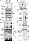
PELI1 deficiency protects against necroptosis. (A) WT and Peli1−/− MEFs were pretreated with Nec-1s or DMSO for 1 h and then treated with TNFα + SM164 + zVAD for indicated periods of time. (B) WT and Peli1−/− MEFs were pretreated with Nec-1s or DMSO for 1 h and then treated with TNFα + 5z7 + zVAD (T5z7Z) or TNFα 10 ng/mL + SM164 + zVAD (TSZ) for 6 h. (C) WT and Peli1−/− MEFs were pretreated with Nec-1s or DMSO for 1 h and then treated with TNFα + 5z7 + zVAD for 6, 12, or 24 h. (D) WT and Peli1−/− MEFs were pretreated with Nec-1s or DMSO for 1 h and then treated with TNFα + SM164 + zVAD for 6, 12, or 24 h. (F and G) HT29 cells were transfected with shRNA targeting Peli1 or Scrambled shRNA for 7 d and then treated with TNFα + SM164 + zVAD 25 (TSZ) or TNFα + CHX + zVAD (TCZ) for 24 h. The cell survival was measured with CellTiterGlo. (E and G) WT and Peli1−/− MEFs (E) or the control and Peli1 knockdown HT29 cells (G) were lysed with SDS buffer and the samples were analyzed by Western blotting with indicated antibodies (G). Concentrations of compounds used Nec-1s, 20 μM; TNFα, 10 ng/mL; SM164, 50 nM (A, B, D, and F), 500 nM (C); zVAD.fmk, 25 μM; CHX, 1 μg/mL.
Fig. 2.
Fig. 2.
PELI1 is indispensable for the formation of RIPK1-RIPK3 necrosome. (A and B) WT or Peli1−/− MEFs were pretreated with zVAD and SM164 for 30 min and then treated with TNFα (TSZ) for indicated periods of time. The cells were lysed with Nonidet P-40 buffer. The cell lysates were immunoprecipitated with anti-RIPK3 antibody (A) or anti-FADD antibody (B). (B) The Nonidet P-40 insoluble fraction was further extracted with 6M urea buffer. The whole-cell lysates and precipitated proteins were analyzed by Western blotting with indicated antibodies. (C) The cells were treated with same condition as A. The cells were lysed with Nonidet P-40 buffer without NEM and analyzed by Western blotting with anti-MLKL in nonreducing SDS/PAGE. (D) WT or Peli1−/− MEFs were pretreated with zVAD and SM164 for 30 min and then treated with TNFα 10 ng/mL (TSZ) for another 2 h. The cells were lysed with 3M urea buffer and immunoprecipitated with anti-K63 ubiquitin ab, or with 6M urea buffer and immunoprecipitated with anti-M1 ubiquitin antibody. (E) WT and Peli1−/− MEFs were treated with FLAG-TNFα 50 ng/mL for indicated periods of time and then lysed with Nonidet P-40 buffer. The cell lysates were immunoprecipitated with anti-FLAG antibody-conjugated agarose (first IP). The immunoprecipitated proteins were eluted with 6M urea buffer. The secondary immunoprecipitation (second IP) of eluted proteins with anti-M1 ubiquitin ab was performed in 6M urea; alternatively, the eluted proteins were diluted into 3M urea buffer and immunoprecipitated with anti-K63 ubiquitin antibody. All immunoprecipitated proteins and whole-cell lysates (WCL) were analyzed by Western blotting with indicated antibodies. Concentrations of compounds used: Nec-1s, 20 μM; TNFα, 10 ng/mL; SM164, 50 nM; zVAD.fmk, 25 μM.
Fig. 3.
Fig. 3.
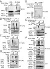
PELI1 is recruited into TNF-RSC in a RIPK1-dependent manner. (A) WT MEFs were treated with FLAG-TNFα for indicated periods of time and then lysed with Nonidet P-40 buffer. The cell lysates were immunoprecipitated with anti-FLAG antibody-conjugated agarose. (B) WT MEFs were pretreated with Nec-1s or vehicle control (0.01% DMSO) for 1 h and then treated with FLAG-TNFα for indicated periods of time. The cells were lysed with Nonidet P-40 buffer, and cell lysates were immunoprecipitated with anti-FLAG antibody-conjugated agarose. (C and D) WT and Ripk1−/− MEFs (C), or WT and Tradd−/− MEFs (D) were treated with FLAG-TNFα for indicated periods of time and then lysed with Nonidet P-40 buffer. The cell lysates were immunoprecipitated with anti-FLAG antibody-conjugated agarose. (E) WT MEFs were pretreated with SM164 or DMSO for 4 h and then treated with FLAG-TNFα for indicated periods of time. The cells were then lysed with Nonidet P-40 buffer, and cell lysates were immunoprecipitated with anti-FLAG antibody-conjugated agarose. (F) WT and Hoip−/− MEFs were treated with FLAG-TNFα for indicated periods of time and then lysed with Nonidet P-40 buffer. The cell lysates were immunoprecipitated with anti-FLAG antibody-conjugated agarose. (G) WT and Abin-1−/− MEFs were treated with FLAG-TNFα for indicated periods of time and then lysed with Nonidet P-40 buffer. The cell lysates were immunoprecipitated with anti-FLAG antibody-conjugated agarose. The whole cell lysates and immunoprecipitated proteins (AF) were analyzed by Western blotting with indicated antibodies. Concentrations of compounds used: FLAG-TNFα, 50 ng/mL; Nec-1s, 20 μM; TNFα, 10 ng/mL; SM164, 50 nM.
Fig. 4.
Fig. 4.
PELI1 mediates K63 ubiquitination of RIPK1 on K115 in a kinase activity-dependent manner. (A) 293T cells were transfected with expression vectors for FLAG-tagged PELI1 and HA-tagged full-length, ΔKD, ΔC, or ΔDD RIPK1 for 24 h and then lysed with Nonidet P-40 buffer. The cell lysates were immunoprecipitated with anti-FLAG antibody-conjugated agarose. (B) 293T cells were pretreated with Nec-1s (20 μM) or vehicle control (0.01% DMSO) for 1 h and then transfected with expression vectors for HA-tagged PELI1 and FLAG-tagged RIPK1 for 24 h. The cells were then lysed with Nonidet P-40 buffer, and cell lysates were immunoprecipitated with anti-HA antibody conjugated agarose. The Nonidet P-40 insoluble fraction was then lysed with 6M urea buffer. (C) 293T cells were transfected with expression vectors for His-K63 ubiquitin and WT or K115R FLAG-RIPK1 with or without that of HA-PELI1 for 24 h and then treated with SM164 or DMSO for 4 h before being lysed with 6M urea. His-tagged proteins were pulled down with Ni-NTA. (D) 293T cells were pretreated with Nec-1s or DMSO for 1 h and then transfected with expression vectors for His-K63 ubiquitin and WT or S161E RIPK1 with or without that of HA-PELI1 for 24 h. The cells were lysed with 6M urea buffer, and His-tagged proteins were pulled down with Ni-NTA. All cell lysates and immunoprecipitated or pulled down proteins were analyzed by Western blotting with indicated antibodies. Concentrations of compounds used: Nec-1s, 20 μM; SM164, 50 nM.
Fig. 5.
Fig. 5.
PELI1 inhibits RIPK1-dependent and RIPK1-independent apoptosis. (A) WT and Peli1−/− MEFs were pretreated with Nec-1s or DMSO for 1 h and then treated with TNFα + 5z7 for the indicated periods. (B) WT and Peli1−/− MEFs were pretreated with Nec-1s or DMSO for 1 h and then treated with TNFα 10 ng/mL + SM164 for the indicated periods. (C) WT and Peli1−/− BV2 cells were pretreated with Nec-1s (20 μM) or DMSO for 1 h and then treated with TNFα 10 ng/mL + 5z7 for the indicated periods. (Right) WT and Peli1−/− BV2 cells were lysed with SDS buffer, and proteins were analyzed by Western blotting with indicated antibodies. (D) WT and Peli1−/− MEFs were pretreated with Nec-1s or DMSO for 1 h and then treated with TNFα + 5z7 or TNFα + CHX for 6 h. (E) WT and Peli1−/− MEFs were pretreated with Nec-1s or DMSO for 1 h and then treated with TNFα + CHX for the indicated periods. The cell survival was determined using CellTiterGlo. Concentrations of compounds used: Nec-1s, 20 μM; TNFα, 10 ng/mL; 5z7, 500 nM; SM164, 50 nM; zVAD.fmk, 25 μM; CHX, 1 μg/mL.
Fig. 6.
Fig. 6.
PELI1 inhibits RIPK1-dependent and RIPK1-independent apoptosis by regulating c-FLIP. (A) WT and Peli1−/− MEFs were treated with TNFα + 5z7 for the indicated periods and then lysed with Nonidet P-40 buffer. The cell lysates were analyzed by Western blotting with the indicated antibodies. (B) WT and Peli1−/− MEFs were treated with TNFα + CHX for the indicated periods and then lysed with Nonidet P-40 buffer. The cell lysates were analyzed by Western blotting with indicated antibodies. (C) The mRNA was extracted from WT and Peli1−/− MEFs and reverse-transcribed into cDNA. The mRNA levels of c-Myc and c-FLIP were determined by quantitative real-time PCR with primers specific to corresponding genes. GAPDH was used as loading control. Concentrations of compounds used: Nec-1s, 20 μM; TNFα, 10 ng/mL; 5z7, 500 nM; CHX, 1 μg/mL.
Fig. 7.
Fig. 7.
Peli1−/− mice are sensitized to TNFα-induced SIRS. (A) Survival curves of nine male WT and nine male Peli1−/− mice (8–12 wk) injected with mTNFα i.v. (mTNFα/mouse = 360 μg/kg). P = 0.0166 according to Gehan-Breslow-Wilcoxon test. (B) Two WT mice and two Peli1−/− mice were killed 6 h after injection with mTNFα and perfused. The spleens from these four mice were lysed and analyzed by Western blotting with indicated antibodies. (C) A model for the role of PELI1 in necroptosis and apoptosis. In necroptotic cells, PELI1 mediates K63 ubiquitination of activated RIPK1 on K115, which can be inhibited by Nec-1s. PELI1-mediated RIPK1 ubiquitination promotes the interaction of RIPK1 and RIPK3 to form necrosome and activation of MLKL to lead to necrosis.

Similar articles

Cited by

References

    1. Christofferson DE, Yuan J. Necroptosis as an alternative form of programmed cell death. Curr Opin Cell Biol. 2010;22:263–268. - PMC - PubMed
    1. Weinlich R, Oberst A, Beere HM, Green DR. Necroptosis in development, inflammation and disease. Nat Rev Mol Cell Biol. 2017;18:127–136. - PubMed
    1. Degterev A, Boyce M, Yuan J. A decade of caspases. Oncogene. 2003;22:8543–8567. - PubMed
    1. Geng J, et al. Regulation of RIPK1 activation by TAK1-mediated phosphorylation dictates apoptosis and necroptosis. Nat Commun. 2017;8:359. - PMC - PubMed
    1. Morioka S, et al. TAK1 kinase switches cell fate from apoptosis to necrosis following TNF stimulation. J Cell Biol. 2014;204:607–623. - PMC - PubMed

Publication types

LinkOut - more resources