Review
. 2016 Apr 11:2:77.
doi: 10.21037/jovs.2016.03.10. eCollection 2016.

Preoperative planning of thoracic surgery with use of three-dimensional reconstruction, rapid prototyping, simulation and virtual navigation

Affiliations
Review

Preoperative planning of thoracic surgery with use of three-dimensional reconstruction, rapid prototyping, simulation and virtual navigation

Samuel Heuts et al. J Vis Surg. .

Abstract

For the past decades, surgeries have become more complex, due to the increasing age of the patient population referred for thoracic surgery, more complex pathology and the emergence of minimally invasive thoracic surgery. Together with the early detection of thoracic disease as a result of innovations in diagnostic possibilities and the paradigm shift to personalized medicine, preoperative planning is becoming an indispensable and crucial aspect of surgery. Several new techniques facilitating this paradigm shift have emerged. Pre-operative marking and staining of lesions are already a widely accepted method of preoperative planning in thoracic surgery. However, three-dimensional (3D) image reconstructions, virtual simulation and rapid prototyping (RP) are still in development phase. These new techniques are expected to become an important part of the standard work-up of patients undergoing thoracic surgery in the future. This review aims at graphically presenting and summarizing these new diagnostic and therapeutic tools.

Keywords: Image-guided surgery; minimally invasive surgery; preoperative planning; thoracic surgery; video-assisted thoracoscopic surgery (VATS).


Conflict of interest statement

Conflicts of Interest: The authors have no conflicts of interest to declare.

Figures

Figure 1
Figure 1
Method for use of the VB navigation system. VB images in the left lower corner, actual bronchoscopy images on the right. The VB images show the target bronchus with a white asterix. VB images adapt automatically to the rotation of the actual bronchoscope [(Reprinted with permission) (7)]. VB, virtual bronchoscopy.
Figure 2
Figure 2
Stepwise approach to diagnose of a (A) ground glass opacity (GGO) found on CT, (B) with a blizzard sign on EBUS, (C) TBB under fluoroscopy guidance, (D) pathology specimen for diagnosis of an adenocarcinoma [(Reprinted with permission) (9)]. CT, computed tomography; EBUS, endobronchial ultrasound; TBB, transbronchial biopsy.
Figure 3
Figure 3
Localization of the tumor in the thorax using 3D CT reconstruction (11). 3D, three-dimensional; CT, computed tomography. Available online: http://www.asvide.com/articles/867
Figure 4
Figure 4
Determination of the affected lung segment and choice for ideal incision level using 3D CT reconstruction (12). 3D, three-dimensional; CT, computed tomography. Available online: http://www.asvide.com/articles/868
Figure 5
Figure 5
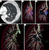
Segmentation of the pulmonary artery, vein and bronchus using 3D CT reconstruction (13). 3D, three-dimensional; CT, computed tomography. Available online: http://www.asvide.com/articles/869
Figure 6
Figure 6
Preoperative simulation of a left upper lobectomy. (A) Confirmation of port placement; (B) status of left upper pulmonary vein, (C,D) pulmonary artery variation [(Reprinted with permission) (14)].
Figure 7
Figure 7
Process of VB-guided bronchoscopic metallic coil placement under fluoroscopy and VATS resection with pathologic specimen examination. (A,B) VB images with actual bronchoscopic images in the right lower corner. Advancement of the bronchoscope into the 6th order bronchus; (C) metallic coil placement under fluoroscopy; (D) CT evaluation of coil placement; (E) intra-operative view of lesion with coil; (F) fluoroscopic view of lesion with coil grasped by pulmonary forceps; (G) macroscopic view of resected lesion with tumor; (H) microscopic finding of the specimen with an adenocarcinoma in situ (magnification, ×400) [(Reprinted with permission) (15)]. CT, computed tomography; VB, virtual bronchoscopy; VATS, video-assisted thoracoscopic surgery.
Figure 8
Figure 8
Concept of iVATS. (A) Preoperative CT depicting a lesion in the right lower lobe; (B) 3D reconstruction showing the relation of the tumor witch the boney structures; (C) pathway planning for T-bar placement; (D) T-bar with fiducial; (E) T-bar placement under fluoroscopy; (F) fluoroscopic view of T-bar placement; (G) intra-operative view of fiducials extending from chest wall to lesion; (H) resected lung specimen [(Reprinted with permission) (18)]. CT, computed tomography; iVATS, image-guided video-assisted thoracoscopic surgery.
Figure 9
Figure 9
A representative case of VAL-MAP. (A) Preoperative CT-images (left) with a 3D image after staining (middle) and post VAL-MAP CT images (right); (B) intra-operative view of the patient with staining [(Reprinted with permission) (19)]. CT, computed tomography; 3D, three-dimensional; VAL-MAP, virtual assisted lung mapping; GGO, ground glass opacity.
Figure 10
Figure 10
Virtual segmentectomy. (A,B) Virtual segmentectomy of S6 with suboptimal resection margins; (C) 3D view of S6 + S8 + S10 a resection; (D) with an optimal resection margin of 25.7 mm; (E) showing segmental bronchi [(Reprinted with permission) (21)]. 3D, three-dimensional.
Figure 11
Figure 11
Creation of 3D pulmonary model. (A-C) Manual segmentation of pulmonary arteries, veins and bronchi by surgeon; (D) depiction of bronchial tree as various sizes of cylinders; (E) 3D pulmonary model [(Reprinted with permission) (22)]. 3D, three-dimensional.
Figure 12
Figure 12
Operating room (OR) scene using binocular stereonavigation. Actual endoscopic 3D display (AED, left) and the binocular stereonavigation display (Navi, right) [(Reprinted with permission) (22)]. 3D, three-dimensional.
Figure 13
Figure 13
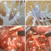
Rapid prototype of the pulmonary vessels and bronchi (A,B) compared to the real-time intra-operative view (C,D) [(Reprinted with permission) (23)].

References

    1. Henschke CI, McCauley DI, Yankelevitz DF, et al. Early lung cancer action project: a summary of the findings on baseline screening. Oncologist 2001;6:147-52. 10.1634/theoncologist.6-2-147 - DOI - PubMed
    1. Petersen RH, Hansen HJ. Learning curve associated with VATS lobectomy. Ann Cardiothorac Surg 2012;1:47-50. - PMC - PubMed
    1. Schimmack S, Hinz U, Wagner A, et al. Maximizing time from the constraining European Working Time Directive (EWTD): The Heidelberg New Working Time Model. Health Econ Rev 2014;4:14. 10.1186/s13561-014-0014-6 - DOI - PMC - PubMed
    1. Niederee MJ, Knudtson JL, Byrnes MC, et al. A survey of residents and faculty regarding work hour limitations in surgical training programs. Arch Surg 2003;138:663-9; discussion 669-71. 10.1001/archsurg.138.6.663 - DOI - PubMed
    1. Reznick RK, MacRae H. Teaching surgical skills--changes in the wind. N Engl J Med 2006;355:2664-9. 10.1056/NEJMra054785 - DOI - PubMed

LinkOut - more resources