. 2017 Oct 27;8(1):239.
doi: 10.1186/s13287-017-0688-x.

Restoring the quantity and quality of elderly human mesenchymal stem cells for autologous cell-based therapies

Affiliations

Restoring the quantity and quality of elderly human mesenchymal stem cells for autologous cell-based therapies

Travis J Block et al. Stem Cell Res Ther. .

Abstract

Background: Degenerative diseases are a major public health concern for the aging population and mesenchymal stem cells (MSCs) have great potential for treating many of these diseases. However, the quantity and quality of MSCs declines with aging, limiting the potential efficacy of autologous MSCs for treating the elderly population.

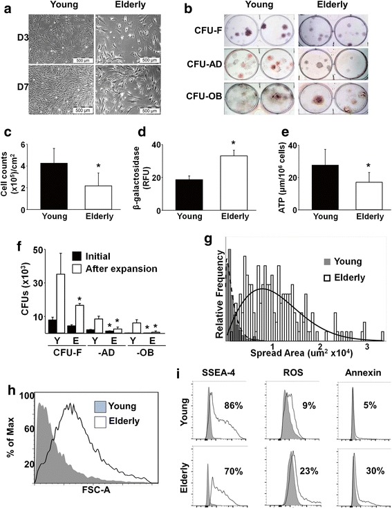
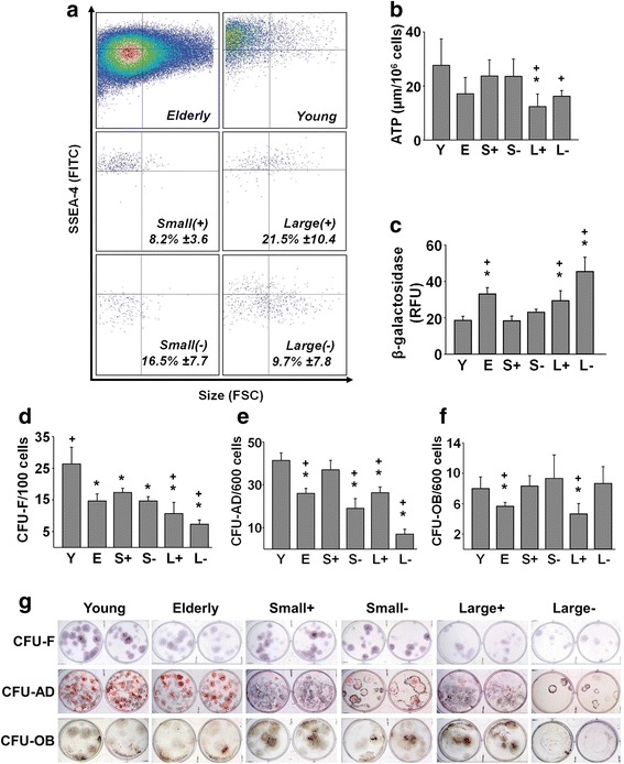
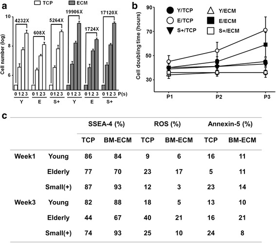
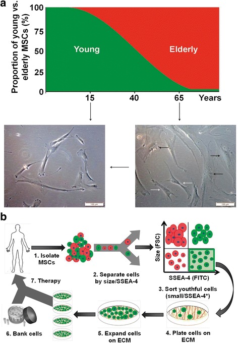
Methods: Human bone marrow (BM)-derived MSCs from young and elderly donors were obtained and characterized using standard cell surface marker criteria (CD73, CD90, CD105) as recommended by the International Society for Cellular Therapy (ISCT). The elderly MSC population was isolated into four subpopulations based on size and stage-specific embryonic antigen-4 (SSEA-4) expression using fluorescence-activated cell sorting (FACS), and subpopulations were compared to the unfractionated young and elderly MSCs using assays that evaluate MSC proliferation, quality, morphology, intracellular reactive oxygen species, β-galactosidase expression, and adenosine triphosphate (ATP) content.

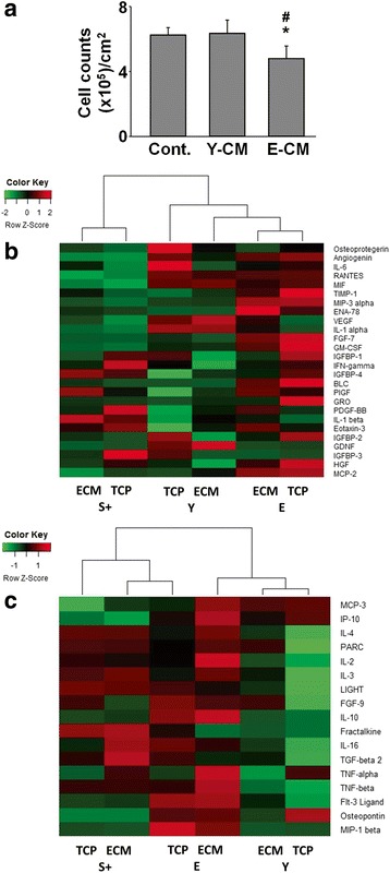
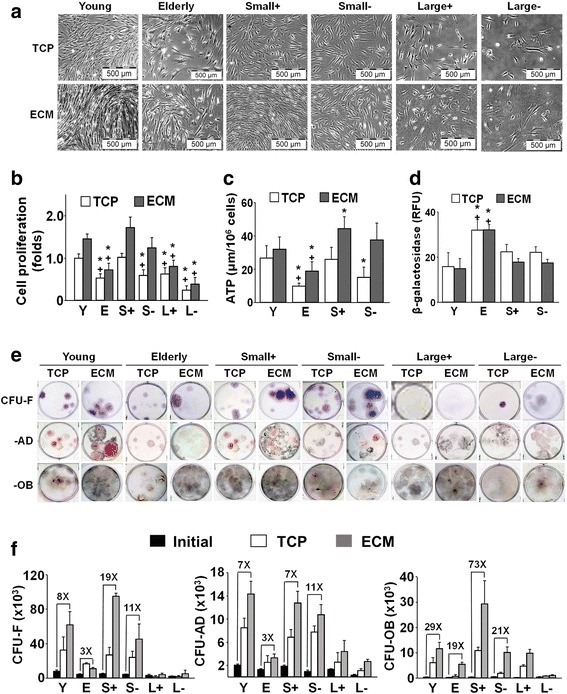
Results: The ISCT-recommended cell surface markers failed to detect any differences between young and elderly MSCs. Here, we report that elderly MSCs were larger in size and displayed substantially higher concentrations of intracellular reactive oxygen species and β-galactosidase expression and lower amounts of ATP and SSEA-4 expression. Based on these findings, cell size and SSEA-4 expression were used to separate the elderly MSCs into four subpopulations by FACS. The original populations (young and elderly MSCs), as well as the four subpopulations, were then characterized before and after culture on tissue culture plastic and BM-derived extracellular matrix (BM-ECM). The small SSEA-4-positive subpopulation representing ~ 8% of the original elderly MSC population exhibited a "youthful" phenotype that was similar to that of young MSCs. The biological activity of this elderly subpopulation was inhibited by senescence-associated factors produced by the unfractionated parent population. After these "youthful" cells were isolated and expanded (three passages) on a "young microenvironment" (i.e., BM-ECM produced by BM cells from young donors), the number of cells increased ≈ 17,000-fold to 3 × 10⁹ cells and retained their "youthful" phenotype.

Conclusions: These results suggest that it is feasible to obtain large numbers of high-quality autologous MSCs from the elderly population and establish personal stem cell banks that will allow serial infusions of "rejuvenated" MSCs for treating age-related diseases.

Keywords: Aging; Cellular senescence; Extracellular matrix; Senescence-associated secretory phenotype; Stem cell markers; Stem cell niche.


Conflict of interest statement

Ethics approval and consent to participate

Bone marrow, obtained from young donors with informed consent and in compliance with all federal regulations, was purchased commercially from LONZA (Walkersville, MD, USA; see company website for additional IRB information: http://www.lonza.com/products-services/bio-research/primary-cells/hematopoietic-cells/unprocessed-bone-marrow.aspx). Bone marrow cells from elderly donors were obtained from consenting patients undergoing total knee or hip arthroplasty according to a protocol approved by the University of Texas Health Science Center at San Antonio (UTHSCSA) Institutional Review Board (IRB) (IRB00000553).

Consent for publication

Not applicable.

Competing interests

X-DC is a board member and shareholder in StemBioSys, Inc. (San Antonio, TX, USA). TJB is currently an employee of StemBioSys, Inc. and receives a salary. X-DC, TJB, and MM are inventors on a pending patent application relating to the content of the manuscript. The remaining authors have no competing or financial interests to declare.

Publisher’s Note

Springer Nature remains neutral with regard to jurisdictional claims in published maps and institutional affiliations.

Figures

Fig. 1
Fig. 1
Compared to young MSCs, elderly MSC quantity and quality is reduced. a, c Brightfield microscopy of MSCs cultured on TCP for 3 or 7 days shows that elderly MSCs were less confluent than young MSCs. By day 7, density of elderly MSCs was significantly lower (n = 16 donors (11 elderly, five young) tested in replicate experiments). b, f After 7 days in culture on TCP, the frequency of young (Y) and elderly (E) MSCs was assessed using CFU-F, CFU-AD, and CFU-OB assays. Cells from elderly donors displayed markedly less CFU replication and differentiation capability (n = 10 donors (five elderly, five young) tested in replicate experiments). d, e β-galactosidase and ATP were measured and elderly MSCs were found to have significantly higher levels of β-galactosidase and significantly lower levels of ATP than young MSCs (n = 10 donors (five elderly, five young) tested in replicate experiments). g, h Cell spread area and cell size (using forward scatter (FSC-A) in flow cytometry) were measured after 3 days in culture. Elderly MSCs cultured on TCP were larger and displayed a wider range of mean cell spread area than young MSCs. i Markers of stemness (SSEA-4) and aging (intracellular ROS and Annexin V) were measured using flow cytometry. Elderly MSCs cultured for 7 days on TCP contained a smaller fraction of cells positive for SSEA-4 and a larger fraction of cells positive for early markers of apoptosis (ROS and Annexin V) than young MSCs (n = 10 donors (five elderly, five young) tested in replicate experiments). *P < 0.05, vs young MSCs. D day, CFU colony forming unit, F fibroblast, AD adipocyte, OB osteoblast, ATP adenosine triphosphate, ROS reactive oxygen species, SSEA-4 stage-specific embryonic antigen-4
Fig. 2
Fig. 2
A subpopulation of elderly MSCs can be isolated using FACS that exhibits a “youthful” phenotype. a Flow cytometry revealed that young MSCs consisted almost exclusively of small, SSEA-4+ (small(+)) cells (top-right panel, upper-left quadrant), while elderly MSCs were more heterogeneous (top-left panel). After sorting elderly MSCs by size (small vs large) and SSEA-4 expression (positive vs negative), four subpopulations were obtained (lower four panels). Mean ± SD of each subpopulation shown; small(+) cells represented on average 8.2% of the elderly MSCs (n = 16 donors (11 elderly, five young) tested in replicate experiments). b, c After isolation, the unfractionated young and elderly MSCs and four subpopulations (S+, S–, L+, L–) were assayed for ATP content and β-galactosidase expression. Compared to elderly MSCs, ATP levels tended to be higher in young MSCs and the small(+) and small(–) subpopulations, but these differences did not achieve statistical significance. In contrast, large(+), but not large(–), cells contained significantly lower levels of ATP than young or small(+) MSCs (P = 0.021). β-galactosidase expression was significantly increased in the elderly MSCs and large(+) and large(–) subpopulations, compared to young and small(+) MSCs, suggesting the presence of senescent cells (n = 10 donors (five elderly, five young) tested in replicate experiments). dg CFU assays (CFU-F, CFU-AD, and CFU-OB) were performed immediately after isolation to determine the enrichment of MSCs. Young MSCs consistently formed more colonies than elderly MSCs in all assays. Small(+) cells were equivalent to young MSCs in the CFU-AD and CFU-OB assays (n = 10 donors (five elderly, five young)). *P < 0.05, vs young MSCs; +P < 0.05, vs small(+) MSCs. S small, L large, Y young, E elderly, RFU relative fluorescence units, CFU colony forming unit, F fibroblast, AD adipocyte, OB osteoblast, ATP adenosine triphosphate, ROS reactive oxygen species, SSEA-4 stage-specific embryonic antigen-4
Fig. 3
Fig. 3
Elderly MSCs produce cytokines, associated with SASP, capable of inhibiting young MSC proliferation. a Conditioned media (CM) were collected from 7-day cultures of young and elderly MSCs on TCP and then added to naïve cultures of young MSCs at a ratio of 1 part CM:2 parts fresh media. Proliferation after 7 days was less in cultures treated with CM from elderly MSCs, suggesting the presence of inhibitory factors (n = 6 donors (three elderly, three young)). b CM from young, small(+), and elderly MSCs cultured for 7 days on TCP or ECM were analyzed using a cytokine array and a heatmap of the SASP-associated cytokines prepared. Small(+) cells expressed less SASP-related cytokines than either young or elderly MSCs. Assays were performed in duplicate using pooled CM (n = 3 donors/group). c CM were collected, analyzed as in (b), and a heatmap of the non-SASP-associated cytokines prepared. Non-SASP-related cytokine production by small(+) cells was similar to that of elderly MSCs, suggesting that some characteristics of the elderly heritage remain. Assays were performed in duplicate using pooled CM (n = 3 donors/group). *P < 0.05, vs CM from young MSCs; #P < 0.05, vs control media. S small, Y young, E elderly, ECM extracellular matrix, TCP tissue culture plastic
Fig. 4
Fig. 4
Expansion of elderly MSC subpopulations on ECM increases cell number and preserves stemness of small(+) cells. a, b Brightfield microscopy of young and elderly MSCs and isolated subpopulations cultured for 7 days on TCP or ECM revealed that growth on ECM significantly enhanced proliferation of young MSCs and both types of small MSCs. Proliferation was calculated as a fold-change by normalizing cell counts at the end of culture to young cells on TCP (n = 16 donors (11 elderly, five young) tested in replicate experiments). c, d ATP levels, but not β-galactosidase expression, were significantly increased in small(+) and small(–) cells and elderly MSCs with culture on ECM (vs TCP) for 7 days, suggesting that ECM promoted maintenance of cell metabolism and inhibited senescence. There were insufficient numbers of large(+)/large(–) cells for assay (n = 10 donors (five elderly, five young) tested in replicate experiments). e, f Following culture on TCP or ECM for 7 days, young and elderly MSCs and isolated subpopulations were detached and seeded at clonal density on TCP for CFU replication assays (CFU-F, CFU-AD, and CFU-OB). The CFU results were consistent with the proliferation data. Culture of young MSCs on ECM significantly enhanced CFU-AD and CFU-OB production, but not CFU-F. Small(+) cells similarly displayed a significant increase in CFU-AD and CFU-OB, as well as CFU-F, production with culture on ECM. CFU replication was calculated by determining the number of CFUs post culture on ECM or TCP and dividing by the number of CFUs produced by the initial population of cells. Fold increases over the initial number of CFUs are shown (n = 10 donors (five elderly, five young) tested in replicate experiments). *P < 0.05, vs young MSCs; +P < 0.05, vs small(+) MSCs. S small, L large, Y young, E elderly, ECM extracellular matrix, TCP tissue culture plastic, ATP adenosine triphosphate, RFU relative fluorescence units, CFU colony forming unit, F fibroblast, AD adipocyte, OB osteoblast
Fig. 5
Fig. 5
Small(+) cells retain their “youthful” phenotype through multiple passages on ECM. Young (Y), elderly (E), and small(+) (S+) MSCs were seeded at 2000 cells/cm2 onto TCP or ECM and subcultured in growth media for 3 weeks. Cells were passaged every 7 days and reseeded at the same density used initially (n = 10 donors (five elderly, five young) tested in replicate experiments). a Total number of cells obtained after culture increased with each passage and was dependent on culture surface and cell type. Compared to culture on TCP, culture on ECM for three passages dramatically increased the number of Y and S+ cells (19,906-fold and 17,120-fold, respectively). As expected, the yield of E cells was much lower. b Cell doubling time was affected by cell type, culture surface, and passage number. At P3, Y and S+ cells had shorter doubling times on TCP than E cells and culture on ECM retained shorter doubling times (S+ < Y < E). c Immunophenotypic properties of Y and S+ cells were maintained through P3. Both Y and S+ cells maintained high expression of SSEA-4 and low levels of ROS and annexin V with culture on ECM. BM-ECM bone marrow-derived extracellular matrix, TCP tissue culture plastic, P passage, ROS reactive oxygen species, SSEA-4 stage-specific embryonic antigen-4
Fig. 6
Fig. 6
Elderly MSCs with a “youthful” phenotype can be expanded on young ECM for cell-based therapies. a Ratio of young to elderly MSCs changes with chronological age. In the elderly, the proportion of young (“youthful”) MSCs has dropped to approximately 8–10% of the total population. This can be clearly seen in the brightfield micrographs. In the young, virtually all cells display a fibroblastic spindle-shaped morphology. With advancing age, cells become larger and spread across the surface (black arrows), but fibroblastic cells can still be found (white arrows). b Our current studies strongly suggest it is possible to rescue these “youthful” cells by separation from the parent MSC population, based on cell size and SSEA-4 expression, and then expand them on a young ECM to bank large quantities of high-quality autologous MSCs for more effective treatment of age-related diseases. ECM extracellular matrix, FSC forward scatter, MSC mesenchymal stem cell, SSEA-4 stage-specific embryonic antigen-4

References

    1. Guralnik J, Fried L, Salive M. Disability as a public health outcome in the aging population. Ann Rev Public Health. 1996;17:25–46. doi: 10.1146/annurev.pu.17.050196.000325. - DOI - PubMed
    1. Powell J. The power of global aging. Aging Int. 2010;35:1–14. doi: 10.1007/s12126-010-9051-6. - DOI
    1. Ho A, Wagner W, Mahlknecht U. Stem cells and aging. EMBO Rep. 2005;6:S35–8. doi: 10.1038/sj.embor.7400436. - DOI - PMC - PubMed
    1. Rando T. Stem cells, aging and the quest for immortality. Nature. 2006;441:1080–6. doi: 10.1038/nature04958. - DOI - PubMed
    1. Trounson A, McDonald C. Stem cell therapies in clinical trials: progress and challenges. Cell Stem Cell. 2015;17:11–22. doi: 10.1016/j.stem.2015.06.007. - DOI - PubMed

MeSH terms