. 2017 Oct 27;7(1):23.
doi: 10.1186/s13395-017-0140-z.

Treatment with the anti-IL-6 receptor antibody attenuates muscular dystrophy via promoting skeletal muscle regeneration in dystrophin-/utrophin-deficient mice

Affiliations

Treatment with the anti-IL-6 receptor antibody attenuates muscular dystrophy via promoting skeletal muscle regeneration in dystrophin-/utrophin-deficient mice

Eiji Wada et al. Skelet Muscle. .

Abstract

Background: Chronic increases in the levels of the inflammatory cytokine interleukin-6 (IL-6) in serum and skeletal muscle are thought to contribute to the progression of muscular dystrophy. Dystrophin/utrophin double-knockout (dKO) mice develop a more severe and progressive muscular dystrophy than the mdx mice, the most common murine model of Duchenne muscular dystrophy (DMD). In particular, dKO mice have smaller body sizes and muscle diameters, and develop progressive kyphosis and fibrosis in skeletal and cardiac muscles. As mdx mice and DMD patients, we found that IL-6 levels in the skeletal muscle were significantly increased in dKO mice. Thus, in this study, we aimed to analyze the effects of IL-6 receptor (IL-6R) blockade on the muscle pathology of dKO mice.

Methods: Male dKO mice were administered an initial injection (200 mg/kg intraperitoneally (i.p.)) of either the anti-IL-6R antibody MR16-1 or an isotype-matched control rat IgG at the age of 14 days, and were then given weekly injections (25 mg/kg i.p.) until 90 days of age.

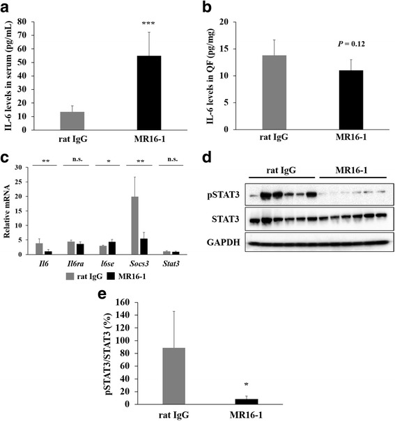
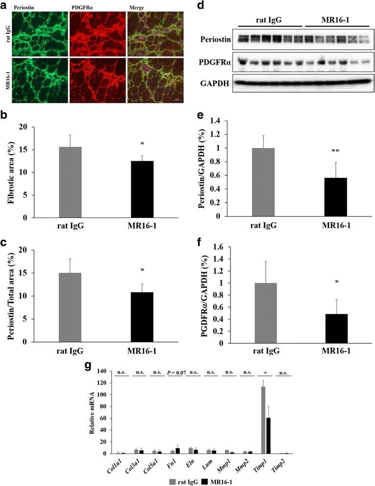
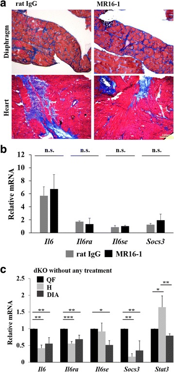
Results: Treatment of dKO mice with the MR16-1 antibody successfully inhibited the IL-6 pathway in the skeletal muscle and resulted in a significant reduction in the expression levels of phosphorylated signal transducer and activator of transcription 3 in the skeletal muscle. Pathologically, a significant increase in the area of embryonic myosin heavy chain-positive myofibers and muscle diameter, and reduced fibrosis in the quadriceps muscle were observed. These results demonstrated the therapeutic effects of IL-6R blockade on promoting muscle regeneration. Consistently, serum creatine kinase levels were decreased. Despite these improvements observed in the limb muscles, degeneration of the diaphragm and cardiac muscles was not ameliorated by the treatment of mice with the MR16-1 antibody.

Conclusion: As no adverse effects of treatment with the MR16-1 antibody were observed, our results indicate that the anti-IL-6R antibody is a potential therapy for muscular dystrophy particularly for promoting skeletal muscle regeneration.

Keywords: Duchenne muscular dystrophy; Fibrosis; Interleukin-6; Muscle regeneration; STAT3.


Conflict of interest statement

Ethics approval

All experimental procedures were approved by the Experimental Animal Care and Use Committees of the National Institute of Neuroscience, National Center of Neurology and Psychiatry, Tokyo Medical University, and the University of Tokyo.

Consent for publication

Not applicable.

Competing interests

The authors declare that they have no competing interests.

Publisher’s Note

Springer Nature remains neutral with regard to jurisdictional claims in published maps and institutional affiliations.

Figures

Fig. 1
Fig. 1
Body weight, KI, serum CK levels, and necrotic area in the quadriceps muscle. a Average body weight of mice at 90 days of age (n = 8 per group). b Radiographs of dKO mice treated with rat IgG or MR16-1. KI was measured using the radiographs. c KI of rat IgG dKO mice (n = 5) and MR16-1-treated dKO mice (n = 8). No significant difference was observed in the average KI between the two groups. d Serum CK levels of the mice at 90 days of age (n = 7–8). e A representative image of anti-mouse IgG-positive necrotic area in the quadriceps muscle. f The percentage of necrotic fibers/total fibers (× 200 magnification). Necrosis was significantly reduced in dKO mice by MR16-1 treatment. *P < 0.05
Fig. 2
Fig. 2
IL-6 levels in serum and the quadriceps muscle. a Serum IL-6 levels of mice at 90 days of age. Serum IL-6 levels of MR16-1-treated dKO mice were increased by about fourfold compared with the rat IgG group. b IL-6 levels of the quadriceps muscle (QF) of dKO mice treated with rat IgG or MR16-1. IL-6 levels were relatively lower in MR16-1-treated dKO mice (P = 0.12). c Quantitative RT-PCR analysis of the levels of Il-6, Il-6ra, Il-6se, Socs3, and Stat3 were measured. All samples were normalized to the expression levels of Lbr. Values of rat IgG and MR16-1 groups were expressed as fold increase of wild-type mice (n = 3–4 per group). d A representative image of Western blot analysis of phosphorylated STAT3 (pSTAT3), total STAT3, and GAPDH. e The expression of pSTAT3 normalized by total STAT3 levels in dKO mice treated with rat IgG or MR16-1 (n = 6 per group). Activated STAT3 levels were significantly decreased in dKO mice by MR16-1 treatment. *P < 0.05, **P < 0.01, ***P < 0.001
Fig. 3
Fig. 3
Histological analysis of the dystrophic phenotypes of the quadriceps muscle. a H&E and Masson’s trichrome staining of the quadriceps muscle from dKO mice. b The percentage of MCIs were counted. c Muscle fibers per field (× 200 magnification) were counted. MR16-1-treated dKO mice increased muscle fibers (P = 0.08). d Histogram of fiber diameters of the rectus femoris muscle (the largest part of the quadriceps). Data are expressed as a percentage of total fiber (n = 8 per group). *P < 0.05
Fig. 4
Fig. 4
Satellite cell content and muscle regeneration. a Pax7 and Ki67 double staining of the quadriceps muscle sections. Arrowheads represent Pax7-positive and Ki67-negative quiescent satellite cells, and arrows represent double-positive (Pax7+/Ki67+) proliferated satellite cells. b The numbers of Pax7+/Ki67+ cells/field and Pax7+/Ki67− cells/field (×400 magnification) in wild-type and dKO mice treated with rat IgG or MR16-1 were compared. The greater number of both activated and quiescent satellite cells was found in dKO mice compared with wild-type mice. While the number of Pax7+/Ki67+ satellite cells was not different, Pax7+/Ki67− satellite cells were significantly increased in MR16-1-treated dKO mice (P < 0.05). c A representative image of eMyHC-positive regenerating muscle fibers in dKO mice treated with rat IgG or MR16-1. d The percentage of eMyHC-positive area per total area. e The number of eMyHC-positive regenerating muscle fibers per field (× 200 magnification). f The percentage of average eMyHC-positive muscle fiber size. g Gene expression levels of myh3, normalized by Lbr, was expressed as fold increase of wild-type mice (n = 3–4 per group). h The percentage of number of myonuclei per fiber. *P < 0.05, **P < 0.01
Fig. 5
Fig. 5
Histological and real-time PCR analyses of inflammatory response in the quadriceps muscle. a F4/80-positive inflammatory area of the quadriceps muscle from dKO mice. b The percentage of F4/80-positive area per total muscle fiber area was quantified. c Gene expression levels of pro-inflammatory cytokines (Il1b, Nos2, Cd68, Tnf, Ifng, and Mcp1, normalized by Lbr) were measured. d Gene expression levels of anti-inflammatory cytokines (Cd163, Il10, Arg1, Mrc1, Fizz1, and Ym1, normalized by Lbr) were measured. The levels were expressed as fold increase of wild-type mice (n = 3–4 per group). *P < 0.05, **P < 0.01
Fig. 6
Fig. 6
The effects of MR16-1 treatment on fibrosis in the quadriceps muscle. a A representative image of double staining of periostin and PDGFRα in the quadriceps muscle. PDGFRα-positive cells were co-localized with periostin-positive fibrotic area. b The quantification of fibrotic area of muscle sections positive for Masson’s trichrome, and c periostin. d Western blot analysis of periostin and PDGFRα expression (n = 6 per group). e The expression levels of periostin and f PDGFRα, normalized by GAPDH, of dKO mice treated with rat IgG or MR16-1 were compared. g The levels of fibrosis-related genes (Col1a1, Col3a1, Col5a1, Fn1, Eln, Lum, Mmp1, Mmp2, Timp1, and Timp2, normalized by Lbr) were measured. The levels were expressed as fold increase of wild-type mice (n = 3–4 per group). *P < 0.05, **P < 0.01
Fig. 7
Fig. 7
IL-6 signaling pathway in the diaphragm and cardiac muscle. a Representative images of H&E and Masson’s trichrome staining of the diaphragm and heart of dKO mice treated with rat IgG or MR16-1. b Gene expression levels of Il-6, Il-6ra, Il-6se, and Socs3 in cardiac muscle were measured by real-time PCR. All samples were normalized using the gene expression levels of Tbp, and expressed as fold increase of wild-type mice (n = 3–4 per group). c The mRNA levels of genes associated with IL-6 (Il-6, Il-6ra, Il-6se, Socs3, and Stat3) were compared among the quadriceps muscle, diaphragm, and cardiac muscle of the same normal (non-treated) dKO mice at 90 days of age (n = 3). *P < 0.05, **P < 0.01, ***P < 0.001

References

    1. Moser H. Duchenne muscular dystrophy: pathogenetic aspects and genetic prevention. Hum Genet. 1984;66:17–40. doi: 10.1007/BF00275183. - DOI - PubMed
    1. Hoffman EP, Brown RH, Kunkel LM. Dystrophin: the protein product of the Duchenne muscular dystrophy locus. Cell. 1987;51:919–928. doi: 10.1016/0092-8674(87)90579-4. - DOI - PubMed
    1. Deconinck N, Dan B. Pathophysiology of duchenne muscular dystrophy: current hypotheses. Pediatr Neurol. 2007;36:1–7. doi: 10.1016/j.pediatrneurol.2006.09.016. - DOI - PubMed
    1. Kharraz Y, Guerra J, Pessina P, Serrano AL, Muñoz-Cánoves P. Understanding the process of fibrosis in Duchenne muscular dystrophy. Biomed Res Int. 2014;2014:965631. doi: 10.1155/2014/965631. - DOI - PMC - PubMed
    1. Mann CJ, Perdiguero E, Kharraz Y, Aguilar S, Pessina P, Serrano AL, Muñoz-Cánoves P. Aberrant repair and fibrosis development in skeletal muscle. Skelet Muscle. 2011;1:21. doi: 10.1186/2044-5040-1-21. - DOI - PMC - PubMed

Publication types

MeSH terms