. 2018 Jan:128:301-313.
doi: 10.1016/j.neuropharm.2017.10.026. Epub 2017 Oct 25.

Functional partnership between mGlu3 and mGlu5 metabotropic glutamate receptors in the central nervous system

Affiliations

Functional partnership between mGlu3 and mGlu5 metabotropic glutamate receptors in the central nervous system

Luisa Di Menna et al. Neuropharmacology. 2018 Jan.

Abstract

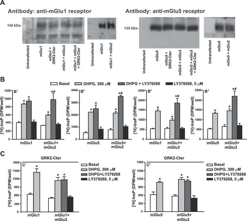
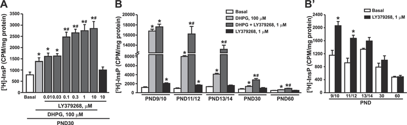
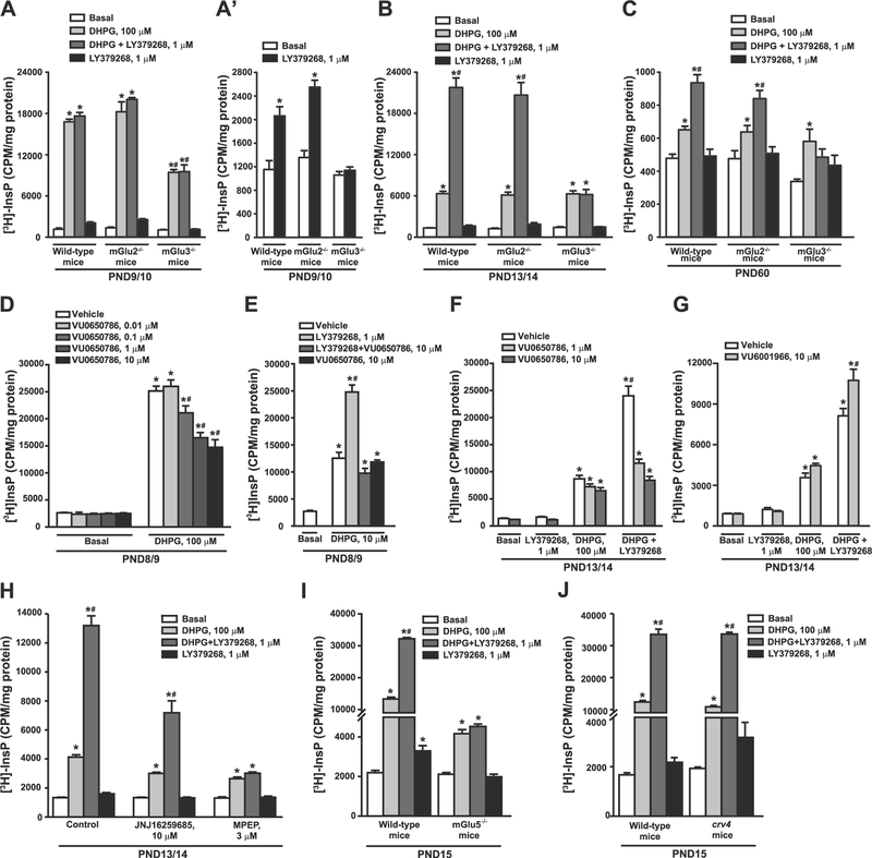
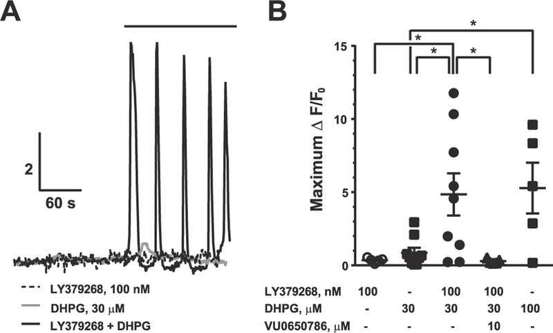
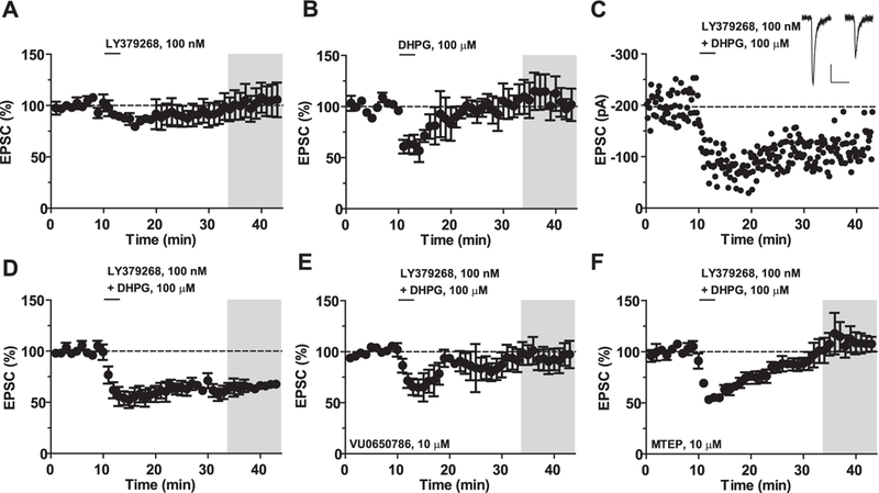
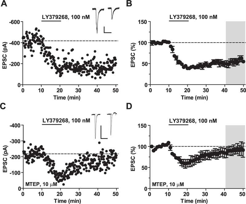
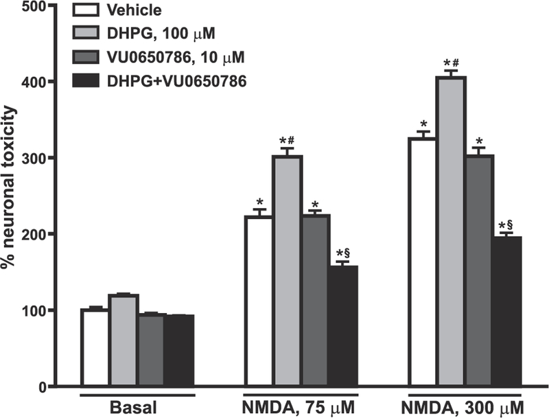
mGlu5 receptors are involved in mechanisms of activity-dependent synaptic plasticity, and are targeted by drugs developed for the treatment of CNS disorders. We report that mGlu3 receptors, which are traditionally linked to the control of neurotransmitter release, support mGlu5 receptor signaling in neurons and largely contribute to the robust mGlu5 receptor-mediated polyphosphoinositide hydrolysis in the early postnatal life. In cortical pyramidal neurons, mGlu3 receptor activation potentiated mGlu5 receptor-mediated somatic Ca2+ mobilization, and mGlu3 receptor-mediated long-term depression in the prefrontal cortex required the endogenous activation of mGlu5 receptors. The interaction between mGlu3 and mGlu5 receptors was also relevant to mechanisms of neuronal toxicity, with mGlu3 receptors shaping the influence of mGlu5 receptors on excitotoxic neuronal death. These findings shed new light into the complex role played by mGlu receptors in physiology and pathology, and suggest reconsideration of some of the current dogmas in the mGlu receptor field.

Keywords: G-protein βγ subunits; Long-term depression; Metabotropic glutamate receptors; Neurodevelopment; Neuronal death; Polyphosphoinositide hydrolysis; Receptor-receptor cross-talk; Synaptic plasticity.


Figures

Fig. 1.
Fig. 1.. Functional cross-talk between group-I and group-II mGlu receptors in transfected HEK 293 cells.
Expression of mGlu1 and mGlu5 receptors in cells transfected with mGlu1 or mGlu5 cDNA in the absence or presence of mGlu2, mGlu3 receptors or GRK2-C-ter cDNA is shown in (A). Stimulation of PI hydrolysis by DHPG and/or LY379268 in cells expressing mGlu1 or mGlu5 receptors with or without mGlu2 or mGlu3 receptors is shown in (B). The effect of GRK2-C-ter on the synergism between mGlu3 and mGlu1 or mGlu5 receptors is shown in (C). In (B) and (C), values are mean ± S.E.M. of 3–9 determinations. p < 0.05 (Two-way ANOVA + Bonferroni test) * vs. the respective basal and # vs. the respective DHPG alone (B 1: F(1,16) = 4.69 for transfection factor and F(3,16) = 43.93 for treatment factor; B2: F(1,16) = 11.39 for transfection factor and F(3,16) = 112.73 for treatment factor; B3: F(1,25) = 2.17 for transfection factor and F(3,25) = 27.00 for treatment factor; B4: F(1,47) = 1.04 for transfection factor and F(3,47) = 41.17 for treatment factor; C1: F(1,25) = 4.89 for transfection factor and F(3,25) = 28.97 for treatment factor; C2: F(3,29) = 30.78 for treatment factor).
Fig. 2.
Fig. 2.. Functional partnership between group-II and group-I mGlu receptors in mouse cortical slices at different developmental ages.
Concentration-dependent amplification of DHPG-stimulated PI hydrolysis by LY379268 in cortical slices of mice at PND30 is shown in (A). The age-dependent profile of DHPG-stimulated PI hydrolysis in the absence or presence of LY379268 is shown in (B). Data with LY379268 alone are highlighted in (B). Values are mean ± S.E.M. of 3–4 determinations. p < 0.05 (One-way ANOVA + Fisher’s LSD in A and B and Student’s t-test in B) vs. the respective basal (*), DHPG (#). (A): F(8,27) = 22.49; (B): F(3,11) = 840.6 (PND9/10); F(3, 8) = 132.27 (PND11/12); F(3,8) = 263.84 (PND13/14); F(3,12) = 29.46 (PND30); F(3,12) = 45.25 (PND60); (B): t(5) = −4.84 (PND9/10); t(4) = −5.32 (PND11/12).
Fig. 3.
Fig. 3.. Functional partnership between native group-I and group-II mGlu receptors is mediated by mGlu3 and mGlu5.
Stimulation of PI hydrolysis by DHPG and/or LY379268 in cortical slices prepared from wild-type, mGlu2−/− and mGlu3−/− mice are shown in (A–C), where wild-type data at PND9/10 and 60 are the same data as in Fig. 2B for comparative purposes. Data with LY379268 at PND9/10 are highlighted in (A). The effect of the mGlu3 receptor NAM, VU0650786, on DHPG/LY379268-stimulated PI hydrolysis is shown in (D–F). The effect of the mGlu2 receptor NAM, VU6001966, on DHPG/LY379268-stimulated PI hydrolysis is shown in (G). The effect of the mGlu1 receptor NAM, JNJ16259685, and the mGlu5 receptor NAM, MPEP, on DHPG/LY379268-stimulated PI hydrolysis is shown in (H), where control data are the same data as in Fig. 2B at PND13/14 for comparative purposes. Stimulation of PI hydrolysis by DHPG/LY379268 in cortical slices from mGlu5−/− or crv4 mice and their wild-type counterparts is shown in (I) and (J), respectively. Values are mean ± S.E.M. of 3–5 determinations. p < 0.05 (Two-way ANOVA + Bonferroni test) * vs. the respective basal values and # vs. the DHPG alone or DHPG + LY379268 values obtained in wild-type and mGlu2−/− mice (A: F(2,33) = 110.73 for genotype factor and F(3,33) = 693.82 for treatment factor; A’: F(2,15) = 36.15 for genotype factor and F(3,15) = 77.46 for treatment factor; B: F(2,30) = 36.33 for genotype factor and F(3,30) = 235.35 for treatment factor; C: F(2,35) = 24.79 for genotype factor and F(2,35) = 40.05 for treatment factor). p < 0.05 (Two-way ANOVA + Bonferroni test) * vs. the respective basal values and # vs. the DHPG values (D: F(1,26) = 913.61 for Basal/DHPG factor and F(4,26) = 13.95 for VU0650786 concentrations factor; E: F(1,14) = 70.67 for Basal/DHPG factor and F(3,14) = 57.33 for drugs factor; F: F(3,31) = 29.60 for drugs factor and F(2,31) = 8.56 for VU0650786 concentrations factor; G: F(3,28) = 234.24 for drugs factor and F(1,28) = 9.89 for VU6001966 concentrations factor; H: F(2,24) = 93.54 for drugs - JNJ16259685 and MPEP - factor and F(3,24) = 284.48 for treatment factor. p < 0.05 (Two-way ANOVA + Bonferroni test) * vs. the respective basal values and # vs. the respective DHPG alone values (I: F(1,32) = 2826.74 for genotype factor and F(3,32) = 1763.58 for treatment factor; J: F(1,22) = 160.79 for genotype factor and F(3,22) = 236.83 for treatment factor).
Fig. 4.
Fig. 4.. mGlu 3 receptor activation enhances mGlu5 receptor-mediated somatic Ca2+ mobilization in pyramidal cells of the mouse PFC.
(A) Representative calcium imaging fluorescence experiments. To isolate neuron-autonomous, mGlu5 receptor-specific calcium responses, all experiments were performed in the presence of tetrodotoxin and the selective mGlu1 receptor NAM, VU0490650 (10 μM). Pyramidal cells were loaded with the cell-impermeable fluorescent calcium indicator Fluo-4. (B) Summary of calcium imaging experiments. Values are mean ± S.E.M. *p < 0.05, **p < 0.01, Bonferroni post-tests.
Fig. 5.
Fig. 5.. mGlu3 and mGlu5 receptors synergize to induce LTD in the mouse PFC.
(A) Application of the mGlu2/3 receptor agonist LY379268 (100 nM) for 3 min does not generate a long-term change in the amplitude of the excitatory postsynaptic current (EPSC) (102 ± 14% baseline, n = 4). (B) Application of the mGlu1/5 receptor agonist DHPG (100 μM) for 3 min does not alter long-term excitatory transmission (108 ± 15% baseline, n = 4). (C) Representative experiment showing LTD induced by the combined bath application of LY379268 and DHPG. Inset, representative EPSC traces before and after LTD. Scale bars denote 50 ms and 50 pA. (D) Summary time course of LTD experiments (65 ± 4% baseline, n = 5). (E) VU0650786 blocked the induction of LTD (94 ± 11% baseline, n = 4). (F) MTEP blocked the induction of LTD (100 ± 12% baseline, n = 4). Values are mean ± S.E.M. in A-F.
Fig. 6.
Fig. 6.. Saturating mGlu3 receptor-LTD requires the activation of mGlu5 receptors.
mGlu3 receptor-dependent-LTD requires activation of mGlu5 receptors. (A) Representative experiment displaying LTD of excitatory transmission following bath application of the mGlu2/3 receptor agonist LY379268 (100 nM) for 10 min. Inset, representative excitatory postsynaptic current (EPSC) before and after LTD. All scale bars denote 100 ms and 100 pA. (B) Summary of LTD experiments. LY379268 induces LTD (54 ± 7% baseline, n = 5). (C) Representative experiment following application of the selective mGlu5 receptor NAM, MTEP (10 μM). (D) MTEP blocked the induction of LTD (89 ± 10% baseline, n = 5). Values are mean ± S.E.M. in A-D.
Fig. 7.
Fig. 7.. Amplification of NMDA toxicity by DHPG in rat cortical neurons requires the endogenous activation of mGlu3 receptors.
Pure cultures of rat cortical neurons were challenged with either 875 or 300 μM NMDA for 20 min in the absence or presence of DHPG and/or the mGlu3 receptor NAM, VU0650786. The experiment was carried out in the presence of the mGlu1 receptor NAM, JNJ16259685 (10 μM), to avoid the stimulation of mGlu1 receptors by DHPG. Neuronal toxicity was assessed by trypan blue staining. Values are means ± S.E.M. of 4 determinations. p < 0.05 (One-way ANOVA + Fisher’s LSD) vs. the respective basal values (*), the vehicle values of the same group (NMDA alone) (#), or vs. the respective DHPG values of the same group (§). F(11,36) = 177.91. This experiment was repeated twice and data were confirmed by using the MTT assay.

References

    1. Aghajanian GK, Marek GJ, 2000. Serotonin model of schizophrenia: emerging role of glutamate mechanisms. Brain Res. Brain Res. Rev 31, 302–312. - PubMed
    1. Alagarsamy S, Marino MJ, Rouse ST, Gereau RW 4th, Heinemann SF, Conn PJ, 1999. Activation of NMDA receptors reverses desensitization of mGluR5 in native and recombinant systems. Nat. Neurosci 2, 234–240. - PubMed
    1. Alagarsamy S, Rouse ST, Junge C, Hubert GW, Gutman D, Smith Y, Conn PJ, 2002. NMDA-induced phosphorylation and regulation of mGluR5. Pharmacol. Biochem. Behav 73, 299–306. - PubMed
    1. Alagarsamy S, Saugstad J, Warren L, Mansuy IM, Gereau RW 4th, Conn PJ, 2005. NMDA-induced potentiation of mGluR5 is mediated by activation of protein phosphatase 2B/calcineurin. Neuropharmacology 49 (Suppl. 1), 135–145. - PMC - PubMed
    1. Ango F, Pin JP, Tu JC, Xiao B, Worley PF, Bockaert J, Fagni L, 2000. Dendritic and axonal targeting of type 5 metabotropic glutamate receptor is regulated by homer1 proteins and neuronal excitation. J. Neurosci 20, 8710–8716. - PMC - PubMed

MeSH terms

Substances