. 2017 Oct 27;6(11):e007038.
doi: 10.1161/JAHA.117.007038.

Essentiality of Regulator of G Protein Signaling 6 and Oxidized Ca2+/Calmodulin-Dependent Protein Kinase II in Notch Signaling and Cardiovascular Development

Affiliations

Essentiality of Regulator of G Protein Signaling 6 and Oxidized Ca2+/Calmodulin-Dependent Protein Kinase II in Notch Signaling and Cardiovascular Development

Bandana Chakravarti et al. J Am Heart Assoc. .

Abstract

Background: Congenital heart defects are the most common birth defects worldwide. Although defective Notch signaling is the major cause of mouse embryonic death from cardiovascular defects, how Notch signaling is regulated during embryonic vasculogenesis and heart development is poorly understood.

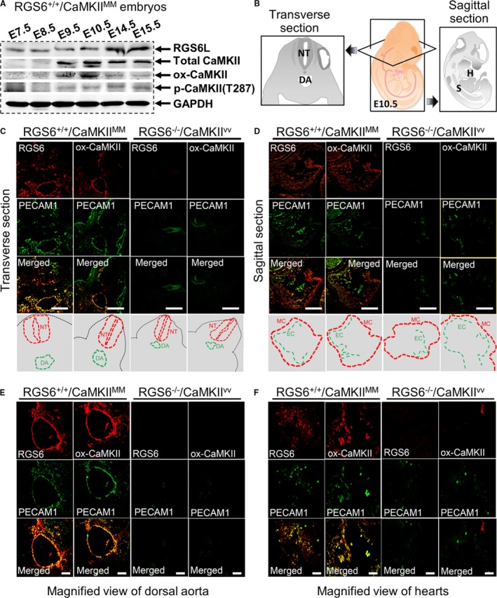
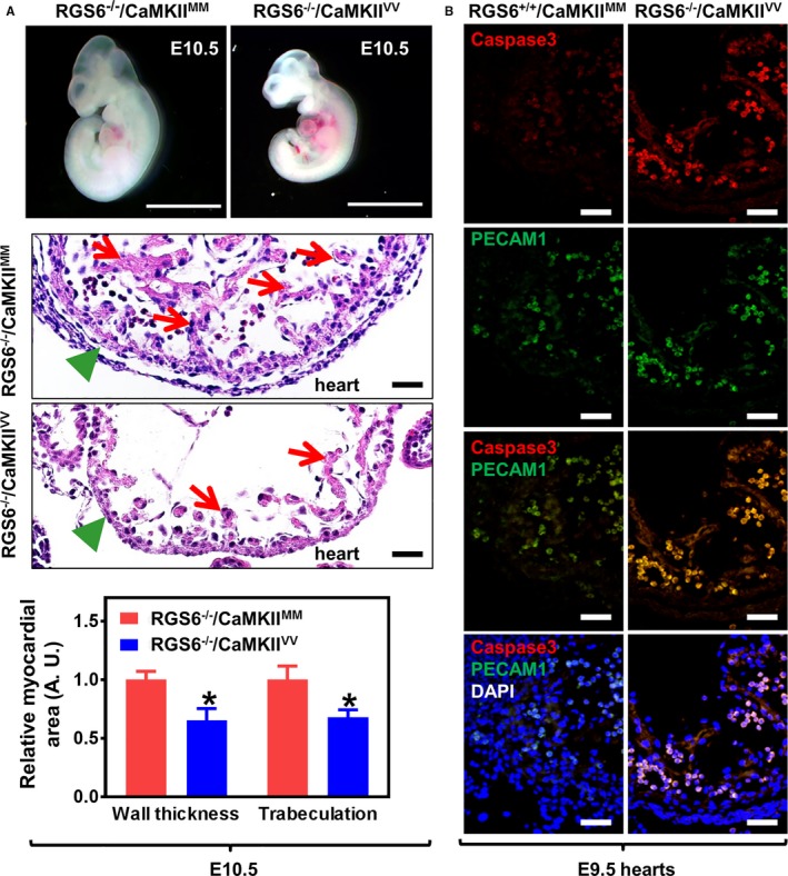
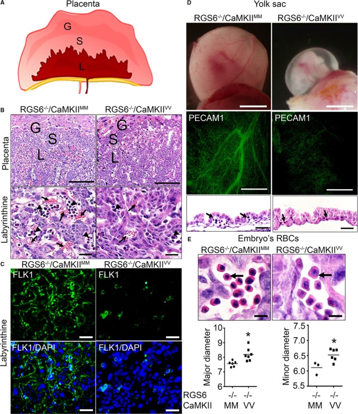
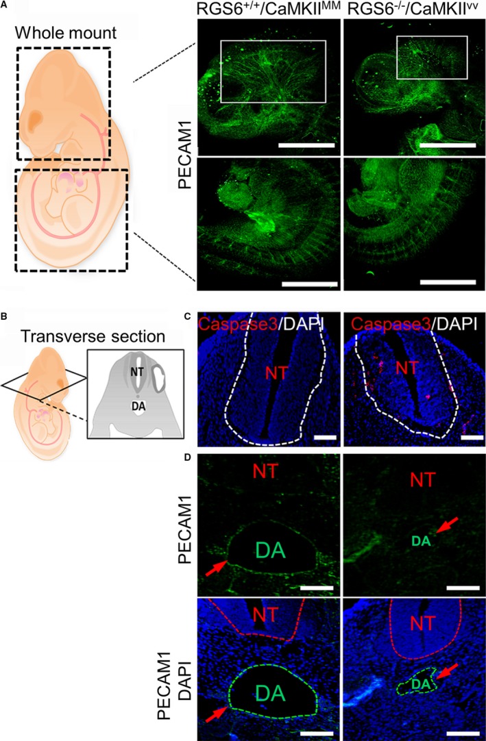
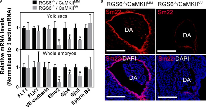
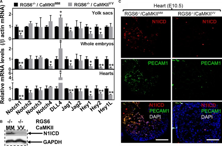
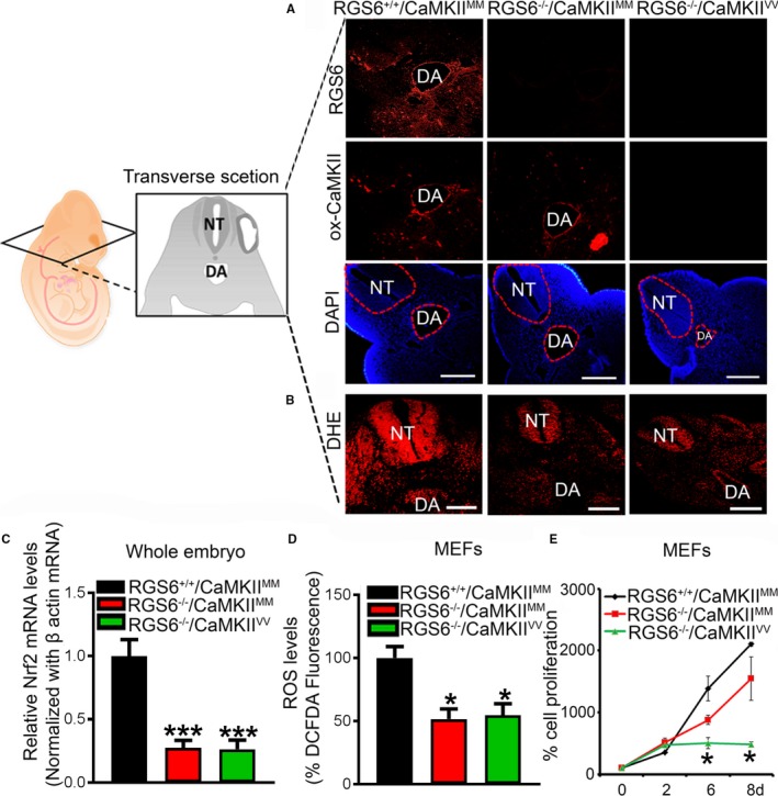
Methods and results: Regulator of G protein signaling 6 (RGS6)-/-/Ca2+/calmodulin-dependent protein kinase II (CaMKII)VV double mutant mice were developed by crossing RGS6-/- mice with mice expressing an oxidation-resistant CaMKIIδ (CaMKIIVV), and the resulting embryonic defects/lethality were investigated using E7.5 to E15.5 embryos. While loss of either RGS6 or oxidized CaMKIIδ does not alter embryogenesis, their combined loss causes defective Notch signaling, severe cardiovascular defects, and embryonic lethality (≈E10.5-11.5). Embryos lacking RGS6 and expressing oxidation-resistant CaMKIIδ exhibit reduced myocardial wall thickness, abnormal trabeculation, and arterial specification defects. Double mutants show vascular remodeling defects, including reduced neurovascularization, delayed neural tube maturation, and small dorsal aortae. These striking cardiovascular defects were accompanied by placental and yolk sac defects in angiogenesis, hematopoiesis, and vascular remodeling similar to what is seen with defective Notch1 signaling. Double mutant hearts, embryos, and yolk sacs exhibit profound downregulation of Notch1, Jagged 1, and Notch downstream target genes Hey1, Hey2, and Hey1L as well as impaired Notch1 signaling in embryos/hearts.

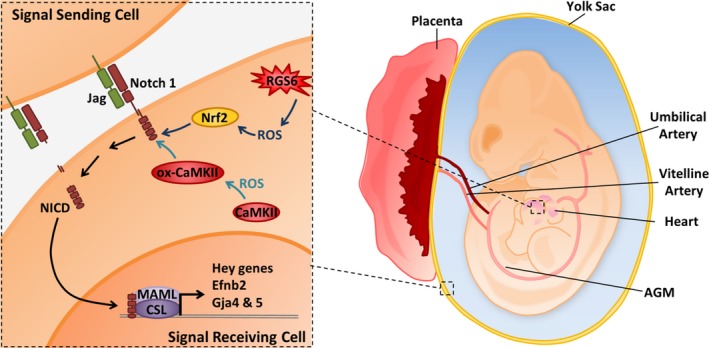
Conclusions: RGS6 and oxidized CaMKIIδ together function as novel critical upstream modulators of Notch signaling required for normal cardiovascular development and embryo survival. Their combined need indicates that they function in parallel pathways needed for Notch1 signaling in yolk sac, placenta and embryos. Thus, dysregulated embryonic RGS6 expression and oxidative activation of CaMKII may potentially contribute to congenital heart defects.

Keywords: Notch signaling; Regulator of G protein signaling 6; cardiovascular development; embryonic lethality; oxidized Ca2+/calmodulin‐dependent protein kinase II.


Figures

Figure 1
Figure 1
Expression of Regulator of G protein signaling 6 (RGS6) and oxidized Ca2+/calmodulin‐dependent protein kinase II (ox‐CaMKII) in mouse embryos. A, Western blot analysis of RGS6, ox‐CaMKII, phosphorylated CaMKII (p‐CaMKII), and total Ca2+/calmodulin‐dependent protein kinase II (CaMKII) expression in RGS6+/+/CaMKIIMM embryos at different gestation periods. B, Schematic illustration of transverse and sagittal sections of embryos used for immunostaining. C, Immunohistochemical analyses of RGS6 and ox‐CaMKII (red) expression in the dorsal aorta of embryos (transverse section) at E10.5 and co‐localization with the endothelial marker platelet endothelial cell adhesion molecule 1 (PECAM1) (green, upper). The schematic view showing the location of the neural tube (NT) and dorsal aorta (DA), in the respective pictures, are indicated by dotted red lines and green lines, respectively (lower). D, Immunohistochemical analysis of RGS6 and ox‐CaMKII (red) expression in the hearts of embryos (sagittal section) at E10.5 and co‐localization with PECAM1 (green, upper). The schematic view showing the location of the myocardium (MC) and endocardium (EC), for the respective pictures, are indicated by dotted red lines and green lines, respectively (lower). E, Co‐localization of RGS6 and ox‐CaMKII (red) with PECAM1 (green) in the DA region at higher magnification from embryo sections at E10.5. F, Co‐localization of RGS6 and ox‐CaMKII (red) with PECAM1 (green) immunostaining of endothelial cells in heart at higher magnification from E10.5 embryo sections. Decreased numbers of positive PECAM1‐stained endothelial cells were found in RGS6−/−/CaMKIIVV embryos compared with RGS6+/+/CaMKIIMM embryos. Scale bars, 50 μm (C and D) and 20 μm (E and F). H indicates heart; S, somites.
Figure 2
Figure 2
Cardiovascular abnormalities in Regulator of G protein signaling 6 (RGS6)−/−/Ca2+/calmodulin‐dependent protein kinase II (CaMKII)VV embryos. A, Representative images of freshly dissected live E10.5 embryos (upper panel, scale bar=200 μm) and hematoxylin and eosin (H&E) staining of heart sections taken from E10.5 embryos (middle panel, scale bar=20 μm). Green arrowheads indicate normal (RGS6−/−/CaMKIIMM) and thinned (RGS6−/−/CaMKIIVV) myocardium and red arrows indicate regions of normal trabeculation in RGS6−/−/CaMKIIMM hearts and its loss in RGS6−/−/CaMKIIVV hearts of H&E‐stained sections (middle panel). Both myocardial thickness and the level of trabeculation were significantly reduced in RGS6−/−/CaMKIIVV embryos when compared with RGS6−/−/CaMKIIMM embryos, analyzed by Mann–Whitney test (lower panel). N=7 to 9 embryos, *P<0.05. B, Increased caspase 3 (red) and platelet endothelial cell adhesion molecule 1 (PECAM1) (green)–stained double‐positive cells (yellow) in the heart ventricle of RGS6−/−/CaMKIIVV embryos at E9.5 compared with RGS6−/−/CaMKIIMM. Scale bar=20 μm. DAPI indicates 4′,6‐diamidino‐2‐phenylindole.
Figure 3
Figure 3
Vascular remodeling and hematopoietic defects in Regulator of G protein signaling 6 (RGS6)−/−/Ca2+/calmodulin‐dependent protein kinase II (CaMKII)VV placentas, yolk sacs, and embryos. A, Schematic representation of different layers of placenta in mice; giant (G), spongiotrophoblast (S), and labyrinthine (L) layers. B, Hematoxylin and eosin (H&E) staining of RGS6−/−/CaMKIIMM and RGS6−/−/CaMKIIVV placenta illustrating G, S, and L layers. Higher magnification images of the labyrinthine layer reveals reduced maternal and fetal blood–filled vasculature in RGS6−/−/CaMKIIVV placentas. Arrows indicate maternal blood (anucleated) and arrowheads indicate fetal blood (nucleated). Scale bars=200 μm (placenta) and 5 μm (L). C, Fetal liver kinase 1 immunostaining of the placental L region revealed a reduction in embryonic vessels in RGS6−/−/CaMKIIVV placentas compared with RGS6−/−/CaMKIIMM placentas. Scale bars=50 μm. D, Representative images of embryos within their yolk sacs show what appears to be an absence of blood‐filled large vitelline vessels, which we later confirmed using whole‐mount platelet endothelial cell adhesion molecule 1 (PECAM1) immunostaining. H&E staining of yolk sacs section reveals fewer blood/hematopoietic cells in vessels (black arrows) in RGS6−/−/CaMKIIVV yolk sacs at E10.5 compared with RGS6−/−/CaMKIIMM yolk sacs, suggesting altered vascular remodeling and hematopoiesis from blood islands. Scale bars=200 μm (whole‐mount and PECAM1) and 20 μm (yolk sac), and (E) larger red blood cell (RBC) nuclei and cell sizes were observed in RGS6−/−/CaMKIIVV embryo sections. Scale bar=20 μm. Both the major and minor diameter of RBCs were significantly larger in RGS6−/−/CaMKIIVV embryos compared with RGS6−/−/CaMKIIMM embryos analyzed by Mann–Whitney statistical methods. N=3, *P<0.05. DAPI indicates 4′,6‐diamidino‐2‐phenylindole.
Figure 4
Figure 4
Vascular remodeling defects in Regulator of G protein signaling 6 (RGS6)−/−/Ca2+/calmodulin‐dependent protein kinase II (CaMKII)VV embryos. A, Schematic view of embryos represents the head and tail region used for whole‐mount platelet endothelial cell adhesion molecule 1 (PECAM1) immunostaining. Results from whole‐mount PECAM1 immunostaining of embryos revealed a lack of large blood vessels within the fetal brain region (indicated by white rectangles) of RGS6−/−/CaMKIIVV compared with RGS6−/−/CaMKIIMM embryos, although no defects on intersomitic vessels in the tail region of embryos were observed. Scale bars=200 μm. B, Schematic view of transverse section of embryos used for immunostaining (C and D). RGS6−/−/CaMKIIVV embryos show a developmentally delayed neural tube (smaller in size) (C), which may be caused by increased cell death, as represented by the caspase 3–positive cells. Scale bars=20 μm. D, Embryo sections showed a reduction in the size of the dorsal aorta and a thin endothelial layer around the dorsal aorta as indicated by red arrows in RGS6−/−/CaMKIIVV relative to RGS6−/−/CaMKIIMM embryos. The dotted lines in 4′,6‐diamidino‐2‐phenylindole (DAPI) staining indicate different regions as shown by abbreviation; NT (neural tube), and DA (dorsal aorta). Scale bars=20 μm.
Figure 5
Figure 5
Arterial differentiation and maturation defects in Regulator of G protein signaling 6 (RGS6)−/−/Ca2+/calmodulin‐dependent protein kinase II (CaMKII)VV embryos. A, mRNA expression levels of endothelial precursors (fms related tyrosine kinase 1 [FLT1] and fetal liver kinase 1 [FLK1]), the vascular endothelial marker (VE‐cadherin), arterial markers (Ephrin B2, gap junction protein alpha 4, and gap junction protein alpha 5), and the venous marker (EphrinB4) were analyzed by quantitative polymerase chain reaction in E10.5 embryos and their yolk sacs. N=3, *P<0.001. B, Sm22 immunostaining of the dorsal aorta revealed a lack of smooth muscle recruitment in RGS6−/−/CaMKIIVV embryos compared with RGS6−/−/CaMKIIMM embryos. Scale bars=20 μm.
Figure 6
Figure 6
Impaired Notch signaling in Regulator of G protein signaling 6 (RGS6)−/−/Ca2+/calmodulin‐dependent protein kinase II (CaMKII)VV embryos. A, quantitative polymerase chain reaction indicated that expression of the Notch receptor (Notch1), Notch ligand (Jagged [Jag] 1), and the Notch downstream target genes Hey1 and Hey2 were decreased in RGS6−/−/CaMKIIVV yolk sacs, embryos, and hearts relative to RGS6−/−/CaMKIIMM embryos. In contrast, Hey1L was only decreased in embryos and heart in double mutants N=3, *P<0.05, **P<0.01 RGS6−/−/CaMKIIVV vs RGS6−/−/CaMKIIMM. B, Western blot analysis indicated that Notch1 intracellular domain (N1ICD) expression was reduced in RGS6−/−/CaMKIIVV embryos compared with RGS6−/−/CaMKIIMM embryos. C, Immunohistochemistry using N1ICD antibody showed decreased expression levels of N1ICD levels in the heart endothelial layer of RGS6−/−/CaMKIIVV embryos compared with RGS6−/−/CaMKIIMM embryos as Notch intracellular domain (red) co‐localizes with platelet endothelial cell adhesion molecule 1 (PECAM1) (green). Scale bars=50 μm. DAPI indicates 4′,6‐diamidino‐2‐phenylindole.
Figure 7
Figure 7
Regulator of G protein signaling 6 (RGS6), oxidized Ca2+/calmodulin‐dependent protein kinase II (ox‐CaMKII), and reactive oxygen species (ROS) expression in embryos of different genotypes. A, Detection of RGS6 (red) and ox‐CaMKII (red) levels in wild‐type (RGS6+/+/CaMKIIMM), RGS6−/− (RGS6−/−/Ca2+/calmodulin‐dependent protein kinase II [CaMKII]MM), and RGS6−/−/CaMKIIVV double mutant embryos by immunostaining. Scale bars=50 μm. B, Effects of combined loss of RGS6 and ox‐CaMKII on superoxide (red) generation in the head region of embryos as shown by dihydroethidium (DHE) staining. The transverse sectioning plane of embryos is represented in the schematic diagram. Scale bars=50 μm. C, Relative Nrf2 mRNA expression levels were analyzed by quantitative polymerase chain reaction in RGS6+/+/CaMKIIMM, RGS6−/−/CaMKIIMM, and RGS6−/−/CaMKIIVV embryos. D, ROS levels in mouse embryonic fibroblasts (MEFs) using 2′,7′‐dichlorodihydrofluorescein diacetate fluorescence. E, Cell proliferation of MEFs at different days using MTT assay. N=3. *P<0.05, ***P<0.01 RGS6−/−/CaMKIIMM or RGS6−/− CaMKIIVV vs RGS6+/+/CaMKIIMM. DA indicates dorsal aorta; DAPI, 4′,6‐diamidino‐2‐phenylindole; NT, neural tube.
Figure 8
Figure 8
Schematic illustration of Regulator of G protein signaling 6 (RGS6)– and/or Ca2+/calmodulin‐dependent protein kinase II (CaMKII)–dependent activation of Notch signaling in embryonic heart and vasculature. In this article we provide evidence that RGS6 and oxidated CaMKII (ox‐CaMKII) function in parallel pathways as critical upstream modulators of Notch signaling. RGS6‐mediated reactive oxygen species (ROS) generation modulates Nrf2 levels while RGS6‐independent ROS oxidizes CaMKII to control Notch signaling for proper cardiovascular development. We hypothesize that together, RGS6 and ox‐CaMKII in signal‐receiving cells (receptor‐ expressing cells), which promote Notch1 receptor and ligand (Jagged [Jag]) interaction and culminate in the proteolytic cleavage of Notch and the release of the Notch intracellular domain (NICD) into the cytoplasm, NICD translocation into the nucleus, and NICD‐mediated gene transcription upon association with other transcriptional regulators CBF1/Su(H)/Lag‐1, CSL, and mastermind‐like (MAML). However, it is possible that RGS6 and ox‐CaMKII are also present in signal‐sending cells (ligand‐expressing cells) and work in a similar manner. In addition, the parallel pathways utilized by RGS6 and ox‐CaMKII (including their respective ROS; RGS6‐dependent ROS [moderate‐high level] and non‐RGS6‐ROS [basal‐low level]) are denoted using different shades of blue to emphasize their separate and independent functions. AGM indicates aorta‐gonad‐mesonephros (contains the dorsal aorta); Efnb2, Ephrin B2.

References

    1. Hoyert DL, Mathews TJ, Menacker F, Strobino DM, Guyer B. Annual summary of vital statistics: 2004. Pediatrics. 2006;117:168–183. - PubMed
    1. Bruneau BG. The developmental genetics of congenital heart disease. Nature. 2008;451:943–948. - PubMed
    1. Niessen K, Karsan A. Notch signaling in cardiac development. Circ Res. 2008;102:1169–1181. - PubMed
    1. Brand T. Heart development: molecular insights into cardiac specification and early morphogenesis. Dev Biol. 2003;258:1–19. - PubMed
    1. Marcelo KL, Goldie LC, Hirschi KK. Regulation of endothelial cell differentiation and specification. Circ Res. 2013;112:1272–1287. - PMC - PubMed

MeSH terms