

Erythropoietin inhibits osteoblast function in myelodysplastic syndromes via the canonical Wnt pathway
- PMID: 29079596
- PMCID: PMC5777191
- DOI: 10.3324/haematol.2017.172726
Erythropoietin inhibits osteoblast function in myelodysplastic syndromes via the canonical Wnt pathway
Abstract
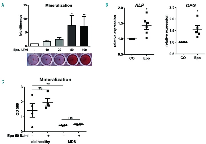
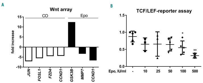
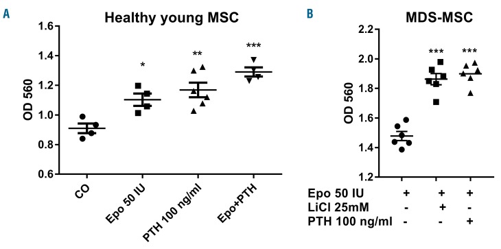
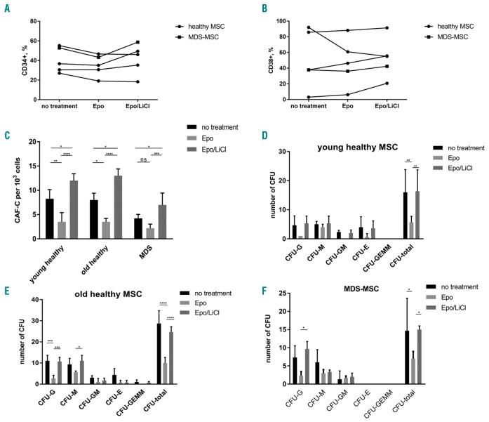
The effects of erythropoietin on osteoblasts and bone formation are controversial. Since patients with myelodysplastic syndromes often display excessively high erythropoietin levels, we aimed to analyze the effect of erythropoietin on osteoblast function in myelodysplastic syndromes and define the role of Wnt signaling in this process. Expression of osteoblast-specific genes and subsequent osteoblast mineralization was increased in mesenchymal stromal cells from healthy young donors by in vitro erythropoietin treatment. However, erythropoietin failed to increase osteoblast mineralization in old healthy donors and in patients with myelodysplasia, whereas the basal differentiation potential of the latter was already significantly reduced compared to that of age-matched controls (P<0.01). This was accompanied by a significantly reduced expression of genes of the canonical Wnt pathway. Treatment of these cells with erythropoietin further inhibited the canonical Wnt pathway. Exposure of murine cells (C2C12) to erythropoietin also produced a dose-dependent inhibition of TCF/LEF promoter activity (maximum at 500 IU/mL, -2.8-fold; P<0.01). The decreased differentiation capacity of erythropoietin-pretreated mesenchymal stromal cells from patients with myelodysplasia could be restored by activating the Wnt pathway using lithium chloride or parathyroid hormone. Its hematopoiesis-supporting capacity was reduced, while reactivation of the canonical Wnt pathway in mesenchymal stromal cells could reverse this effect. Thus, these data demonstrate that erythropoietin modulates components of the osteo-hematopoietic niche in a context-dependent manner being anabolic in young, but catabolic in mature bone cells. Targeting the Wnt pathway in patients with myelodysplastic syndromes may be an appealing strategy to promote the functional capacity of the osteo-hematopoietic niche.
Copyright© 2018 Ferrata Storti Foundation.
Figures
Similar articles
-
Cajanolactone A from Cajanus cajan Promoted Osteoblast Differentiation in Human Bone Marrow Mesenchymal Stem Cells via Stimulating Wnt/LRP5/β-Catenin Signaling.Molecules. 2019 Jan 12;24(2):271. doi: 10.3390/molecules24020271. Molecules. 2019. PMID: 30642055 Free PMC article.
-
Effects of rigosertib on the osteo-hematopoietic niche in myelodysplastic syndromes.Ann Hematol. 2019 Sep;98(9):2063-2072. doi: 10.1007/s00277-019-03756-1. Epub 2019 Jul 16. Ann Hematol. 2019. PMID: 31312928
-
Transforming growth factor β1-mediated functional inhibition of mesenchymal stromal cells in myelodysplastic syndromes and acute myeloid leukemia.Haematologica. 2018 Sep;103(9):1462-1471. doi: 10.3324/haematol.2017.186734. Epub 2018 May 17. Haematologica. 2018. PMID: 29773599 Free PMC article.
-
The importance of WNT pathways for bone metabolism and their regulation by implant topography.Eur Cell Mater. 2012 Jul 12;24:46-59. doi: 10.22203/ecm.v024a04. Eur Cell Mater. 2012. PMID: 22791372 Review.
-
In vitro biology of the hematopoietic system in myelodysplastic syndromes.Cancer Treat Res. 2001;108:25-43. doi: 10.1007/978-1-4615-1463-3_2. Cancer Treat Res. 2001. PMID: 11702602 Review. No abstract available.
Cited by
-
Erythropoietin enhances osteogenic differentiation of human periodontal ligament stem cells via Wnt/β-catenin signaling pathway.Drug Des Devel Ther. 2019 Jul 26;13:2543-2552. doi: 10.2147/DDDT.S214116. eCollection 2019. Drug Des Devel Ther. 2019. PMID: 31440036 Free PMC article.
-
Machine Learning of Bone Marrow Histopathology Identifies Genetic and Clinical Determinants in Patients with MDS.Blood Cancer Discov. 2021 Mar 22;2(3):238-249. doi: 10.1158/2643-3230.BCD-20-0162. eCollection 2021 May. Blood Cancer Discov. 2021. PMID: 34661156 Free PMC article.
-
Effects of Erythropoietin-Promoted Fracture Healing on Bone Turnover Markers in Cats.J Funct Biomater. 2024 Apr 17;15(4):106. doi: 10.3390/jfb15040106. J Funct Biomater. 2024. PMID: 38667563 Free PMC article.
-
Active hematopoiesis triggers exosomal release of PRDX2 that promotes osteoclast formation.Physiol Rep. 2021 Feb;9(3):e14745. doi: 10.14814/phy2.14745. Physiol Rep. 2021. PMID: 33587325 Free PMC article.
-
Effects of Erythropoietin in White Adipose Tissue and Bone Microenvironment.Front Cell Dev Biol. 2020 Nov 24;8:584696. doi: 10.3389/fcell.2020.584696. eCollection 2020. Front Cell Dev Biol. 2020. PMID: 33330462 Free PMC article. Review.
References
-
- Ogunshola OO, Bogdanova AY. Epo and non-hematopoietic cells: what do we know? Methods Mol Biol. 2013;982:13–41. - PubMed
-
- Rolfing JH, Baatrup A, Stiehler M, Jensen J, Lysdahl H, Bunger C. The osteogenic effect of erythropoietin on human mesenchymal stromal cells is dose-dependent and involves non-hematopoietic receptors and multiple intracellular signaling pathways. Stem Cell Rev. 2014;10(1):69–78. - PubMed
Publication types
MeSH terms
Substances
LinkOut - more resources
Full Text Sources
Other Literature Sources
Medical
