. 2018 Mar 21;38(12):3001-3012.
doi: 10.1523/JNEUROSCI.2460-17.2017. Epub 2017 Oct 27.

Basolateral Amygdala Neurons Maintain Aversive Emotional Salience

Affiliations

Basolateral Amygdala Neurons Maintain Aversive Emotional Salience

Auntora Sengupta et al. J Neurosci. .

Abstract

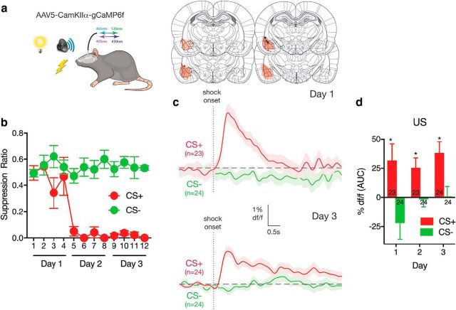
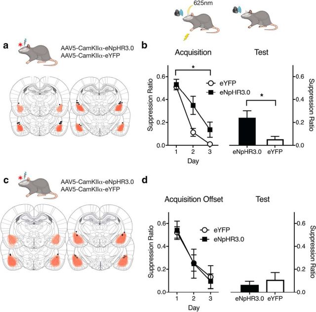
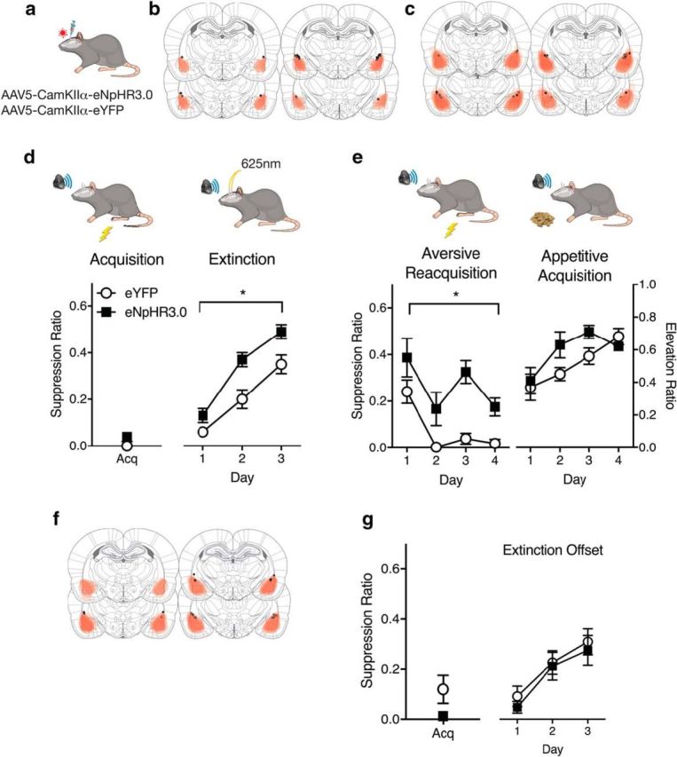
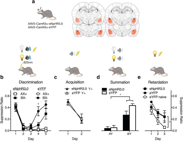
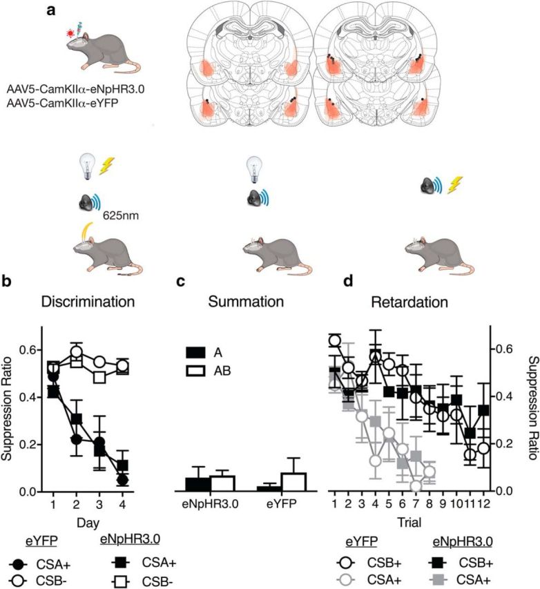
BLA neurons serve a well-accepted role in fear conditioning and fear extinction. However, the specific learning processes related to their activity at different times during learning remain poorly understood. We addressed this using behavioral tasks isolating distinct aspects of fear learning in male rats. We show that brief optogenetic inhibition of BLA neurons around moments of aversive reinforcement or nonreinforcement causes reductions in the salience of conditioned stimuli, rendering these stimuli less able to be learned about and less able to control fear or safety behaviors. This salience reduction was stimulus-specific, long-lasting, and specific to learning about, or responding to, the same aversive outcome, precisely the goals of therapeutic interventions in human anxiety disorders. Our findings identify a core learning process disrupted by brief BLA optogenetic inhibition. They show that a primary function of the unconditioned stimulus-evoked activity of BLA neurons is to maintain the salience of conditioned stimuli that precede it. This maintenance of salience is a necessary precursor for these stimuli to gain and maintain control over fear and safety behavior.SIGNIFICANCE STATEMENT The amygdala is essential for learning to fear and learning to reduce fear. However, the specific roles served by activity of different amygdala neurons at different times during learning is poorly understood. We used behavioral tasks isolating distinct aspects of learning in rats to show that brief optogenetic inhibition of BLA neurons around moments of reinforcement or nonreinforcement disrupts maintenance of conditioned stimulus salience. This causes a stimulus-specific and long-lasting deficit in the ability of the conditioned stimulus to be learned about or control fear responses. These consequences are the precisely goals of therapeutic interventions in human anxiety disorders. Our findings identify a core learning process disrupted by brief BLA optogenetic inhibition.

Keywords: amygdala; conditioning; fear; salience.


Figures

Figure 1.
Figure 1.
Fiber photometry of BLA neurons during fear learning. a, AAV-CaMKIIα-gCaMP6f was applied to BLA. AAV expression across all animals, with each rat represented at 10% opacity. Black circles represent the ventral tip of the fiber optic cannulae. b, Rats received differential fear conditioning (CS shock, CS+; CS no shock, CS) and learned to fear CS+ but not CS. c, Ca2+ transients during shock US delivery on CS+ trials and omission on CS trials on days 1 and 3 of conditioning. n indicates number of trials. d, Areas under the ΔF/F curve for the 5 s from onset of shock or shock omission across conditioning. n indicates number of trials. n = 6. *p < .05.
Figure 2.
Figure 2.
Characterization of BLA neurons. a, Typical voltage response of eNpHR3.0-positive neurons to positive and negative current injections. b, Voltage responses to photoinhibition of various durations. Bars represent timing of the stimuli. c, Action potential train evoked by brief current injections (5 ms, 10 Hz) plotted above the response to a 7 s light pulse delivered during the AP train, and the response to a 7 s light pulse in the absence of depolarizing current injections. d, Response to synaptic stimulation (top), synaptic stimulation during photoinhibition (middle), and photoinhibition alone (bottom). n = 7 cells.
Figure 3.
Figure 3.
BLA neurons and fear learning. a, AAV expression across all animals, with each rat represented at 10% opacity. Black circles represent the ventral tip of the fiber optic cannulae. b, eYFP and eNpHR3.0 groups received CS shock pairings with photoinhibition during the shock US. Photoinhibition impaired fear conditioning. A suppression ratio of 0.5 indicates no fear, and a suppression ratio of 0 indicates high fear. c, AAV expression across offset control animals, with each rat represented at 10% opacity. d, Control eYFP and eNpHr3.0 groups received CS shock pairings and photoinhibition randomly during the ITI. Data are mean ± SEM. Group sizes were as follows: eYFP, n = 7; eNpHR3.0, n = 7; eYFP-Offset, n = 7; eNpHR3.0-Offset, n = 5. *p < 0.05.
Figure 4.
Figure 4.
BLA neurons and fear extinction learning. a, AAV-CaMKIIα-eYFP or eNpHr3.0 was applied to BLA. b, AAV expression for animals receiving fear conditioning, extinction, and fear reacquisition, with each rat represented at 10% opacity. Black circles represent the ventral tip of the fiber optic cannulae. c, AAV expression for animals receiving fear conditioning, extinction, and appetitive conditioning, with each rat represented at 10% opacity. d, eYFP (n = 14) and eNpHR3.0 (n = 11) groups received extinction training with photoinhibition during shock omission. Photoinhibition augmented fear loss during extinction. A suppression ratio of 0.5 indicates no fear, and a suppression ratio of 0 indicates high fear. e, eYFP and eNpHr3.0 groups were retrained in fear or appetitive conditioning and showed selective impairments in fear relearning. For aversive conditioning, a suppression ratio of 0.5 indicates no fear, and a suppression ratio of 0 indicates high fear. For appetitive conditioning, an elevation ratio >0.5 indicates CS-elicited magazine entries. f, AAV-CaMKIIα-eYFP or eNpHr3.0 was applied to BLA. AAV expression across all animals, with each rat represented at 10% opacity. Black circles represent the ventral tip of the fiber optic cannulae. g, Control eYFP (n = 7) and eNpHr3.0 (n = 5) groups received extinction training with photoinhibition randomly during the ITI. Photoinhibition had no effect on extinction learning. Data are mean ± SEM. *p < 0.05.
Figure 5.
Figure 5.
BLA neurons and safety learning. a, AAV-CaMKIIα-eYFP or eNpHr3.0 was applied to BLA. AAV expression, with each rat represented at 10% opacity. Black circles represent the ventral tip of the fiber optic cannulae. b, Rats received AX+/BX discrimination training to establish B as a conditioned inhibitor of fear. Photoinhibition during US omission on BX trials slowed discrimination learning. c, Rats then received CS Y+ pairings with shock. d, Summation test showing poorer conditioned inhibition in the eNpHR3.0 group. e, Retardation test showing normal retardation in the eNpHR3.0 group. Data are mean ± SEM. eYFP, n = 7; eNpHR3.0, n = 6. *p < 0.05.
Figure 6.
Figure 6.
BLA neurons and CS preexposure. a, AAV-CaMKIIα-eYFP or eNpHr3.0 was applied to BLA. AAV expression, with each rat represented at 10% opacity. Black circles represent the ventral tip of the fiber optic cannulae. b, Rats received A+/B discrimination training with photoinhibition on B trials. c, Summation test showing preexposure did not transform B into a conditioned inhibitor. d, Retardation test showing normal retardation in both eYFP and eNpHR3.0 groups. Data are mean ± SEM. eYFP, n = 5; eNpHR3.0, n = 5.

Similar articles

Cited by

References

    1. Annau Z, Kamin LJ (1961) The conditioned emotional response as a function of the intensity of the US. J Comp Physiol Psychol 54:428–432. 10.1037/h0042199 - DOI - PubMed
    1. Bevins RA, Ayres JJ (1991) Two issues in Pavlovian fear conditioning: selective fear of bright vs dark, and CS determinants of CR form. Behav Process 24:211–218. 10.1016/0376-6357(91)90076-C - DOI - PubMed
    1. Beyeler A, Namburi P, Glober GF, Simonnet C, Calhoon GG, Conyers GF, Luck R, Wildes CP, Tye KM (2016) Divergent routing of positive and negative information from the amygdala during memory retrieval. Neuron 90:348–361. 10.1016/j.neuron.2016.03.004 - DOI - PMC - PubMed
    1. Davis M. (1992) The role of the amygdala in fear and anxiety. Annu Rev Neurosci 15:353–375. 10.1146/annurev.ne.15.030192.002033 - DOI - PubMed
    1. Ehrlich I, Humeau Y, Grenier F, Ciocchi S, Herry C, Lüthi A (2009) Amygdala inhibitory circuits and the control of fear memory. Neuron 62:757–771. 10.1016/j.neuron.2009.05.026 - DOI - PubMed

Publication types

LinkOut - more resources