. 2017 Oct 27;7(1):14261.
doi: 10.1038/s41598-017-14496-5.

In Vitro Seamless Stack Enzymatic Assembly of DNA Molecules Based on a Strategy Involving Splicing of Restriction Sites

Affiliations

In Vitro Seamless Stack Enzymatic Assembly of DNA Molecules Based on a Strategy Involving Splicing of Restriction Sites

Dong Yu et al. Sci Rep. .

Abstract

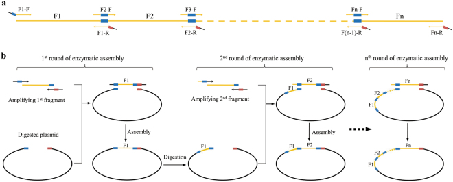
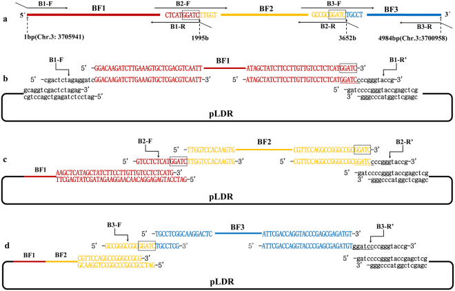
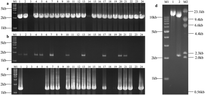
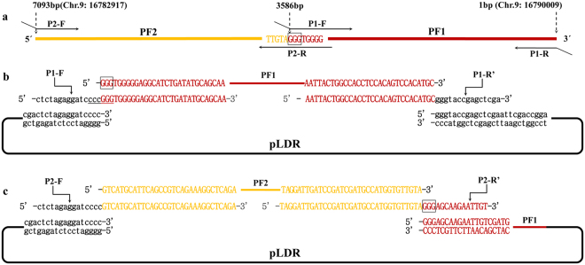
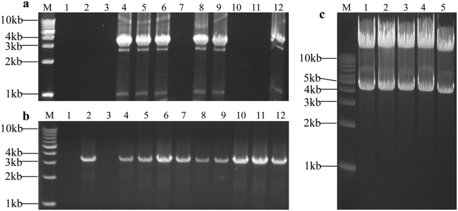
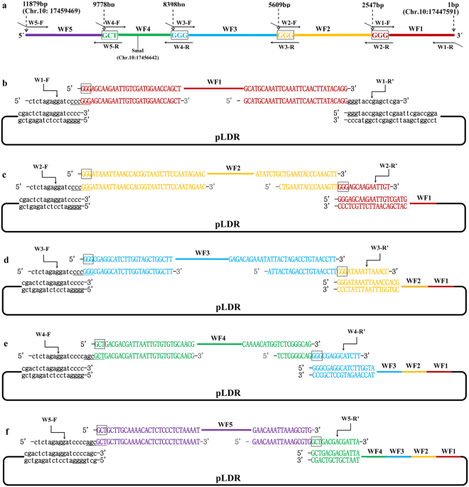
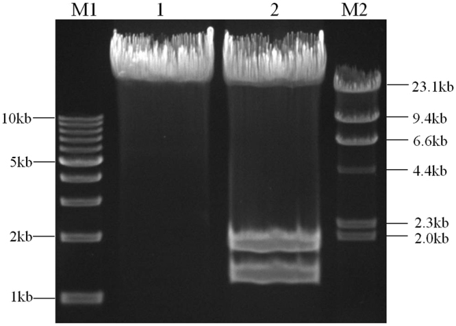
The standard binary enzymatic assembly, which operates by inserting one DNA fragment into a plasmid, has a higher assembly success rate than the polynary enzymatic assembly, which inserts two or more fragments into the plasmid. However, it often leaves a nucleotide scar at the junction site. When a large DNA molecule is assembled stepwise into a backbone plasmid in a random piecewise manner, the scars will damage the structure of the original DNA sequence in the final assembled plasmids. Here, we propose an in vitro Seamless Stack Enzymatic Assembly (SSEA) method, a novel binary enzymatic assembly method involving a seamless strategy of splicing restriction sites via a stepwise process of multiple enzymatic reactions that does not leave nucleotide scars at the junction sites. We have demonstrated the success and versatility of this method through the assembly of 1) a 4.98 kb DNA molecule in the 5' → 3' direction using BamHI to generate the sticky end of the assembly entrance, 2) a 7.09 kb DNA molecule in the 3' → 5' direction using SmaI to generate the blunt end of the assembly entrance, and 3) an 11.88 kb DNA molecule by changing the assembly entrance.


Conflict of interest statement

The authors declare that they have no competing interests.

Figures

Figure 1
Figure 1
Schematic diagram illustrating the design of SSEA. The black line represents pLDR and orange represents the assembled DNA sequence. The thick short line shown in blue and red represents the 2 stitching sites which could be spliced together to regenerate a restriction site. (a) The assembled DNA sequence was divided into several fragments by stitching sites and the orange arrows represent the matching primers with overlapping. (b) After each round of PCR amplification, digestion, and assembly, the original restriction site is regenerated only once in the construct, which then serves as the assembly entrance for the next fragment. By iteration of these steps, a long DNA sequence can be cloned into a plasmid with high fidelity, but without introducing scar sequences.
Figure 2
Figure 2
Assembly of a 4.98 kb DNA molecule by generating the sticky end of the assembly entrance. The stitching sites GGATC are boxed. The line and lowercase sequence shown in black represents pLDR. The line and uppercase sequence shown in red represents the first fragment of BF1, orange represents the second fragment of BF2, and blue represents the third fragment of BF3. (a) The 4.98 kb DNA molecule was divided into 3 fragments by 2 stitching sites GGATC and the matching overlapping primers. (b) The first round of the enzymatic assembly reactions for inserting BF1 is shown. The backbone vector of pLDR was digested by BamHI. The BamHI site was then eliminated in B1-F, and a BamHI site (GGATCc) was restored by splicing the first stitching site and overlapping region in B1-R′ (B1-R′ was a reverse complement with B1-R). When the BF1 amplified with primers of B1-F and B1-R was assembled into pLDR, there was only 1 BamHI site in the assembled products, which could be cut to generate the assembly entrance for the second round. (c) The second round of the enzymatic assembly reaction for inserting BF2 is shown. The intermediate plasmid of pLDR-BF1(4) was digested by BamHI. There was a BamHI site (GGATCc) spliced by the second stitching site and overlapping region in B2-R′ (B2-R′ was a reverse complement with B2-R), but there was no BamHI site spliced in B2-F despite the first stitching site GGATC existing in B2-F. As for the first stitching site GGATC in B2-F, the BF2 must join together with the BF1 seamlessly, and then there is only 1 BamHI site in the assembled products, which could be cut to generate the assembly entrance for the third round. (d) The third round of the enzymatic assembly reaction for inserting BF3 is shown. The intermediate plasmid of pLDR-BF1 ~ BF2(1) was digested by BamHI. As for the second stitching site GGATC in B3-F, the BF3 must join together with the BF2 seamlessly. A BamHI site was retained in the overlapping region of B3-R′ for other consequent applications (B3-R′ was a reverse complement with B3-R).
Figure 3
Figure 3
Identification of the assembly products. M1: TAKARA 1 kb DNA ladder; M2: TAKARA λ-HindIII digest. (a) Plasmids of pLDR-BF1(1)-(24) screened by PCR amplification directly from 24 colonies with primers B1-F and B1-R; the predicted band was 2025 bp. (b) Plasmids of pLDR-BF1~BF2(1)-(24) screened by PCR amplification directly from 24 colonies with the primers B2-F and B2-R; the predicted band was 1687 bp. (c) Plasmids of pLDR-BF1~BF3(1)-(24) screened by PCR amplification directly from 24 colonies with the primers B3-F and B3-R; the predicted band was 1362 bp. (d) 1: The final vector of pLDR-BF1~BF3 digested by BamHI; the predicted band was 14777 bp. 2: The final vector of pLDR-BF1~BF3 digested by SalI; the predicted bands were 12629 bp and 2148 bp.
Figure 4
Figure 4
Assembly of a 7.09 kb DNA molecule by generating the blunt end of the assembly entrance. The stitching sites GGG are boxed. The black line and lowercase sequence represents pLDR. The red line and uppercase sequence represents the first fragment of PF1, and orange represents the second fragment of PF2. (a) The 7.09 kb DNA molecule was divided into two fragments by 1 stitching site GGG and the matching overlapping primers. (b) The first round of enzymatic assembly reactions for inserting PF1 is shown. The backbone vector of pLDR was digested by SmaI. The SmaI site was then eliminated in the forward primer of P1-R′ (P1-R′ was a reverse complement with P1-R), and a SmaI site (cccGGG) was restored by splicing the first stitching site and overlapping region in the reverse primer of P1-F. When the first fragment of PF1 obtained by PCR with primers of P1-F and P1-R was assembled into pLDR, there was only 1 SmaI site in the first round of the assembled products, which could be cut to generate the assembly entrance for the second round. (c) The second round of the enzymatic assembly reaction for inserting PF2 is shown. The intermediate plasmid of pLDR-PF1 was digested by SmaI. As for the first stitching site GGG in P2-R′ (P2-R′ was a reverse complement with P2-R), the second fragment of PF2 must join together with the first fragment of PF1 seamlessly.
Figure 5
Figure 5
Identification of the assembly products. M: TAKARA 1 kb DNA ladder. (a) Plasmids of pLDR-PF1(1)–(12) screened by PCR amplification directly from 12 colonies with primers P1-F and P1-R; the predicted band was 3616 bp. (b) Plasmids of pLDR-PF1 ~ PF2(1)–(12) screened by PCR amplification directly from 12 colonies with the primers P2-F and P2-R; the predicted band was 3537 bp. (c) Digestion tests for the final vector of pLDR-PF1 ~ PF2. Five final plasmid DNAs analyzed by restriction digestion with KpnI; the predicted bands were 12615 bp and 4271 bp.
Figure 6
Figure 6
Assembly of the 11.88 kb DNA molecule by changing assembly entrance in the assembly process. The stitching sites GGG and GCT are boxed. The line and sequence shown in black represent pLDR, red represents the first fragment of WF1, orange represents the second fragment of WF2, blue represents the third fragment of WF3, green represents the fourth fragment of WF4, and purple represents the fifth fragment of WF5. The primer of W1-R′ was reverse complement with W1-R, as well as W2- R′ and W2-R, W3- R′ and W3-R, W4- R′ and W4-R, W5- R′ and W5-R. (a) The 11.88 kb DNA molecule was divided into five fragments by three stitching sites GGG and one stitching site GCT. (b) The first round of assembly reaction for inserting WF1. The pLDR was digested by SmaI. When WF1 was assembled into pLDR, there was only 1 SmaI site in the assembly products, which can be cut to generate the assembly entrance for the second round. (c) The second round of assembly reaction for inserting WF2. The pLDR-WF1 was digested by SmaI. As for the first stitching site GGG in W2-R′, WF2 must join together with WF1 seamlessly, and then there is only 1 SmaI site in the second round of assembled products. (d) The third round of assembly reaction for inserting WF3. The pLDR-WF1 ~ WF2 was digested by SmaI. As for the second stitching site GGG in W3-R′, WF3 must join together with WF2 seamlessly, and then there is only 1 SmaI site in the third round of assembled products. (e) The fourth round of assembly reaction for inserting WF4. The pLDR-WF1 ~ WF3 was digested by SmaI. An Eco47III was introduced by adding AGC additionally in the overlapping region of W4-F before the stitching site GCT. As for the third stitching site GGG in W4-R′, WF4 must join together with WF3 seamlessly, and then there is only 1 Eco47III site in the fourth round of assembled products. (f) The fifth round of assembly reaction for inserting WF5. The pLDR-WF1~WF4 was digested by Eco47III. The fifth fragment of WF5 seamlessly joins with WF4.
Figure 7
Figure 7
Identification of the final vector of pLDR-W1F1~WF5. M1: TAKARA 1 kb DNA ladder; M2: TAKARA λ-HindIII digest; 1: the plasmid of pLDR-WF1 ~ WF5 digested by Eco47III; the predicted band was 23505 bp; and 2: the plasmid of pLDR-WF1 ~ WF5 digested by SalI; the predicted bands were 20604 bp, 1715 bp and 1186 bp.

Similar articles

Cited by

References

    1. Ma S, et al. DNA synthesis, assembly and applications in synthetic biology. Curr. Opin. Chem. Biol. 2012;16:260–267. doi: 10.1016/j.cbpa.2012.05.001. - DOI - PMC - PubMed
    1. Zhou J, et al. CasHRA (Cas9-facilitated Homologous Recombination Assembly) method of constructing megabase-sized DNA. Nucleic Acids Res. 2016;44:e124. doi: 10.1093/nar/gkw475. - DOI - PMC - PubMed
    1. Ellis T, et al. DNA assembly for synthetic biology: from parts to pathways and beyond. Integr. Biol. 2011;3:109–118. doi: 10.1039/c0ib00070a. - DOI - PubMed
    1. Zhang L, et al. Tandem assembly of the epothilone biosynthetic gene cluster by in vitro site-specific recombination. Sci. Rep. 2011;1:141. doi: 10.1038/srep00141. - DOI - PMC - PubMed
    1. Weber E, et al. A modular cloning system for standardized assembly of multigene constructs. PloS ONE. 2011;6:e16765. doi: 10.1371/journal.pone.0016765. - DOI - PMC - PubMed

Publication types

LinkOut - more resources