Review
. 2019 Jan;44(1):117-132.
doi: 10.1007/s11064-017-2428-0. Epub 2017 Oct 27.

Glutamate Dehydrogenase, a Complex Enzyme at a Crucial Metabolic Branch Point

Affiliations
Review

Glutamate Dehydrogenase, a Complex Enzyme at a Crucial Metabolic Branch Point

Hong Q Smith et al. Neurochem Res. 2019 Jan.

Abstract

In-vitro, glutamate dehydrogenase (GDH) catalyzes the reversible oxidative deamination of glutamate to α-ketoglutarate (α-KG). GDH is found in all organisms, but in animals is allosterically regulated by a wide array of metabolites. For many years, it was not at all clear why animals required such complex control. Further, in both standard textbooks and some research publications, there has been some controversy as to the directionality of the reaction. Here we review recent work demonstrating that GDH operates mainly in the catabolic direction in-vivo and that the finely tuned network of allosteric regulators allows GDH to meet the varied needs in a wide range of tissues in animals. Finally, we review the progress in using pharmacological agents to activate or inhibit GDH that could impact a wide range of pathologies from insulin disorders to tumor growth.

Keywords: Allostery; Glutamate dehydrogenase; Insulin.


Figures

Fig. 1
Fig. 1
Schematic diagram of glutamate dehydrogenase catalysis. Under high glutamate concentrations, glutamate replaces α-ketoglutarate before reduced coenzyme is released, forming an abortive complex. This is slowly resolved and the next catalytic cycle is started (red box). At lower concentrations of substrate (green box), the abortive complex is not formed and the enzyme is rapidly recycled for the next round of catalysis. The GDH·NADH·aKG and the GDH·NADH·Glu complexes are colored blue and red, respectively, to note that the reduced coenzyme in these complexes can be directly observed as blue and red shifted species in pre-steady state stopped-flow analysis. As also noted in the red box, GTP and ADP stabilize and destabilize this abortive complex, respectively. (Color figure online)
Fig. 2
Fig. 2
Regulation of GDH. The red and green lines represent inhibition and activation, respectively. The compounds shown in blue represent synthetic allosteric regulators. The dashed lines represent the antagonism or agonism between the various allosteric regulators. SCHAD and SIRT4 are not allosteric ligands but rather enzymes that interact with GDH and cause inhibition. (Color figure online)
Fig. 3
Fig. 3
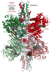
Structure of glutamate dehydrogenase. The image on the left is of the entire GDH hexamer with each subunit shown in different colors. Glutamate (yellow) and NADH (grey) bound to the active site are shown as space filling models. The inhibitor, GTP, is shown as a brick colored space filling model. The images on the right show close up views of GTP and ADP bound to the enzyme. GTP binds between the NAD binding domain and the antenna whereas ADP binds behind the NAD+ binding domain. (Color figure online)
Fig. 4
Fig. 4
Possible location of the leucine activation site. The structure of GDH from Thermus thermophiles complexed with the activator leucine [37] was aligned with bovine GDH. Shown here in mauve is the location of the bound leucine from the bacterial structure to suggest a possible location for the leucine activation site. (Color figure online)
Fig. 5
Fig. 5
Locations of three compounds that inhibit GDH activity. a Shows a wedge of the GDH core, viewed down the threefold axis, with the bound compounds represented as space filling models. Bithionol and GW5074 bind to the same site, between adjacent subunits, and midway between the core of the enzyme and its exterior. Hexachlorophene binds as a ring in the core of the enzyme. b Shows how pairs of GW5074 stack against each other and lie between adjacent GDH subunits. c Shows the aromatic stacking that allows hexachlorophene to form the ring structure in the GDH core
Fig. 6
Fig. 6
The binding location of ECG (and EGCG) on GDH. a Shows the location of bound ECG which is essentially the same as the ADP site. bd Show the effects of mutating residues in contact with ECG on allosteric regulation by ADP, GTP, and EGCG (Figures adapted from [85])
Fig. 7
Fig. 7
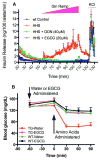
The effect of EGCG on insulin secretion in H454Y transgenic mice β-islets and on the whole animal (Figures adapted from [85]). a TG tissue secretes insulin in response to a Gln ramp stimulation. This is not observed in WT islets, and glutamine-stimulated insulin secretion in TG islets is blocked by the glutaminase inhibitor, DON, and by the GDH inhibitor, EGCG. Note that EGCG, but not DON, brings the basal insulin secretion levels (T = 20 min) down to that of WT (data are mean ± S.E. (error bars), n = 3 for each group). The black line representing WT tissue maybe difficult to see because it lies directly under the TG+EGCG line (green). b This figure shows the effects of oral administration of EGCG on the hypersecretion of insulin in HHS transgenic mice. Plasma glucose levels in WT mice (n = 12 for water- or EGCG-treated mice) are essentially unaffected by oral administration of water or EGCG prior to the administration of the amino acid mixture. However, the plasma glucose levels rapidly drop in the HHS TG mice (n = 12) upon the administration of the amino acid mixture, but this is blocked when the animals are fed EGCG (n = 16) prior to the amino acid challenge. (Color figure online)
Fig. 8
Fig. 8
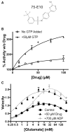
Identification of a new GDH activator using high throughput screening. By screening the compound library with GTP in the assay, N1-[4-(2-aminopyrimidin-4-yl)phenyl]-3-(trifluoromethyl) benzene-1-sulfonamide, Maybridge Hitscreen compound 75-E10, was identified as a promising new activator of GDH (figures adapted from [92]) a The structure of 75-E10. b This figure shows the effects of 75-E10 on GDH in the presence and absence of GTP. As with leucine and ADP, 75-E10 has a small effect on GDH activity alone (~ 50% activation) but a much larger effect when abrogating GTP inhibition (~ 330% activation). c This graph shows that 75-E10 is more effective than the natural activator, ADP, with a > 10-fold better ED50 and activation of the enzyme over a broader range of conditions

Similar articles

Cited by

References

    1. Hudson RC, Daniel RM. L-Glutamate dehydrogenases: distribution, properties and mechanism. Comp Biochem Physiol. 1993;106B:767–792. - PubMed
    1. Frieden C. Glutamate dehydrogenase VI: survey of purine nucleotides and other effects on the enzyme from various sources. J Biol Chem. 1965;240:2028–2037. - PubMed
    1. Frieden C. Glutamic dehydrogenase I. The effect of coenzyme on the sedimentation velocity and kinetic mechanism. J Biol Chem. 1959;234:809–814. - PubMed
    1. Tomkins GM, Yielding KL, Curran JF. The influence of diethylstilbestrol and adenosine diphosphate on pyridine nucleotide coenzyme binding by glutamic dehydrogenase. J Biol Chem. 1962;237:1704–1708. - PubMed
    1. Bailey JS, Bell ET, Bell JE. Regulation of bovine glutamate dehydrogenase. J Biol Chem. 1982;257:5579–5583. - PubMed

MeSH terms