. 2018 Apr;75(7):1269-1284.
doi: 10.1007/s00018-017-2694-7. Epub 2017 Oct 28.

Human endoglin as a potential new partner involved in platelet-endothelium interactions

Affiliations

Human endoglin as a potential new partner involved in platelet-endothelium interactions

Elisa Rossi et al. Cell Mol Life Sci. 2018 Apr.

Abstract

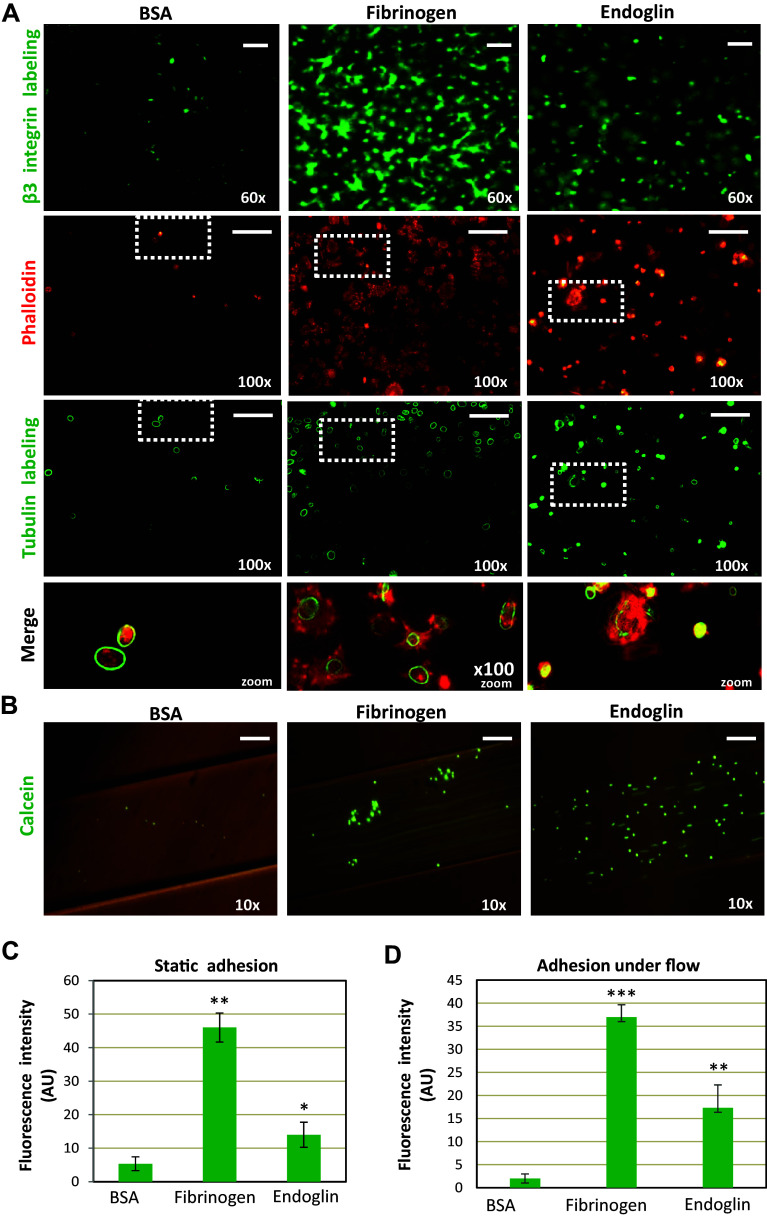
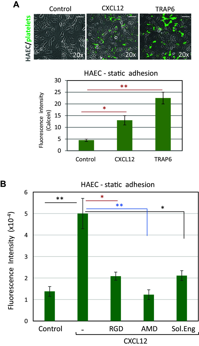
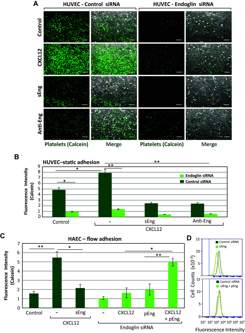
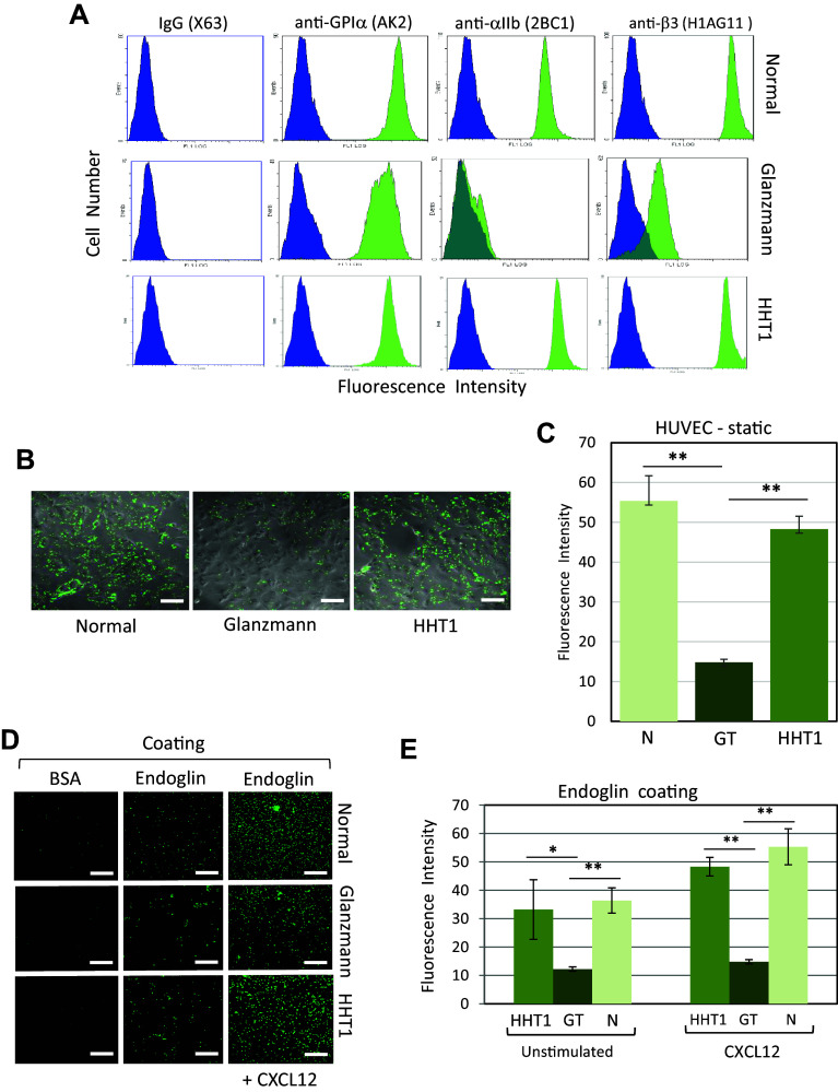
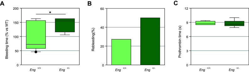
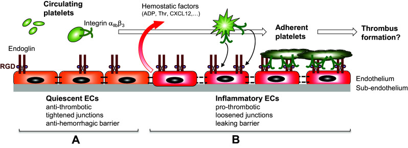
Complex interactions between platelets and activated endothelium occur during the thrombo-inflammatory reaction at sites of vascular injuries and during vascular hemostasis. The endothelial receptor endoglin is involved in inflammation through integrin-mediated leukocyte adhesion and transmigration; and heterozygous mutations in the endoglin gene cause hereditary hemorrhagic telangiectasia type 1. This vascular disease is characterized by a bleeding tendency that is postulated to be a consequence of telangiectasia fragility rather than a platelet defect, since platelets display normal functions in vitro in this condition. Here, we hypothesize that endoglin may act as an adhesion molecule involved in the interaction between endothelial cells and platelets through integrin recognition. We find that the extracellular domain of human endoglin promotes specific platelet adhesion under static conditions and confers resistance of adherent platelets to detachment upon exposure to flow. Also, platelets adhere to confluent endothelial cells in an endoglin-mediated process. Remarkably, Chinese hamster ovary cells ectopically expressing the human αIIbβ3 integrin acquire the capacity to adhere to myoblast transfectants expressing human endoglin, whereas platelets from Glanzmann's thrombasthenia patients lacking the αIIbβ3 integrin are defective for endoglin-dependent adhesion to endothelial cells. Furthermore, the bleeding time, but not the prothrombin time, is significantly prolonged in endoglin-haplodeficient (Eng +/-) mice compared to Eng +/+ animals. These results suggest a new role for endoglin in αIIbβ3 integrin-mediated adhesion of platelets to the endothelium, and may provide a better understanding on the basic cellular mechanisms involved in hemostasis and thrombo-inflammatory events.

Keywords: CXCL12; HHT; Hereditary hemorrhagic telangiectasia; Preeclampsia; RGD; TGF-β.


Conflict of interest statement

The authors declare no competing financial interests.

Figures

Fig. 1
Fig. 1
Platelet adhesion on endoglin-coated wells. a Platelets resuspended in Walsh’s buffer were incubated for 30 min in endoglin-, fibrinogen- or BSA-coated wells. After washing the plates, platelets were labeled with an anti-β3 integrin subunit antibody (green fluorescence), or with phalloidin (red fluorescence) and anti-tubulin antibody (green fluorescence), as described under “Materials and methods”. The zoom of merged phalloidin and tubulin staining is shown in the lower row. Scale bars, 10 µm. Quantification of platelets labeled with the anti-β3 was performed using ImageJ software and is shown in c. Mean values ± SEM of three different preparations of platelets each tested in triplicate are displayed. Significant differences were observed between endoglin or fibrinogen coating and BSA control substrate (**p < 0.005; *p < 0.05). b Platelet adhesion under flow was assessed using the microfluidic system of BioFlux, as described under “Materials and methods”. Calcein-labeled platelets were perfused in flow chambers coated with the indicated proteins, allowed to adhere in the absence of flow for 10 min, and then subjected to 2 dynes/cm2 for 2 min. Platelets bound to the substrate were visualized by microscopy. Scale bars, 60 µm. Quantification of platelet adhesion was performed using ImageJ software and is shown in d. Mean values ± SEM of three different preparations of platelets each tested in triplicate are displayed. Significant differences were observed between endoglin or fibrinogen coating and BSA control substrate (***p < 0.001; **p < 0.005). Of note, due to the different experimental conditions, adhesion values obtained under static (c) or flow conditions (d) are not comparable. AU arbitrary units
Fig. 2
Fig. 2
Effect of CXCL12 and TRAP6 on platelet adhesion to endothelial cells. a Calcein-labeled platelets were incubated for 10 min on HAEC monolayers in the absence or presence of CXCL12 or thrombin receptor activating peptide 6 (TRAP6) and washed twice with PBS. Adhesion of platelets to EC was visualized by fluorescence microscopy (×20 magnification) (top panels). Scale bars, 40 µm. Quantification of platelet adhesion repeated in quadruplicates with two different platelet preparations and analyzed by ImageJ software is shown in the histogram. Mean values ± SEM are displayed. **p < 0.01; *p < 0.05. b Calcein-labeled platelets were incubated for 10 min on HAEC monolayers in the absence (control) or presence of CXCL12, either alone or with the RGD peptide, the CXCR4 inhibitor AMD3100, or the recombinant extracellular part of endoglin (Sol.Eng), as indicated. Platelets were visualized by fluorescence microscopy and quantified (quadruplicates with two different platelet preparations) with ImageJ software. Mean values ± SEM are displayed. **p < 0.005; *p < 0.01
Fig. 3
Fig. 3
Effect of endoglin silencing on adhesion of platelets to EC. a Static adhesion of platelets to HUVEC. Platelets labeled with calcein were incubated for 10 min on HUVEC monolayers, previously treated with a siRNA specific for endoglin or with a scrambled siRNA, as described under “Materials and methods”. Incubation was carried out in the absence or presence of CXCL12 (100 ng/mL), sEng (50 ng/mL), or the anti-endoglin mAb P4A4. After washing twice with PBS, adhesion under static conditions was visualized by fluorescence microscopy (×20 magnification). Scale bars, 40 µm. b Quantification of platelet adhesion shown in a repeated in triplicates with two different platelet preparations and analyzed using ImageJ software. Mean values ± SEM are displayed. **p < 0.005; *p < 0.05. c Platelet adhesion under flow was assessed using a Maastricht Instrumentation equipment. Calcein-labeled platelets were perfused in flow chambers coated with HAEC, previously untreated or treated with siRNA specific for endoglin, in the presence of CXCL12 or sEng, as indicated. Rescue experiments of endoglin expression were performed by nucleofection of HAEC with the endoglin expression vector pCEXV-EndoL (pEng). Platelets were allowed to adhere in the absence of flow for 10 min, and then subjected to 2 dynes/cm2 for 2 min. Quantification of platelet adhesion (triplicates with two different platelet preparations), was performed using ImageJ software. Mean values ± SEM are displayed. **p < 0.005; *p < 0.05. d HAEC were nucleofected with siRNA specific for endoglin (siEng) or with a scrambled siRNA (control siRNA), as indicated. Rescue experiments of endoglin expression were performed by nucleofection of HAEC with the endoglin expression vector pCEXV-EndoL (pEng). The expression levels of endoglin were determined by immunofluorescence flow cytometry using specific antibodies prior to adhesion experiments shown in c. The vertical blue line indicates the fluorescence intensity of HAEC stained with an irrelevant isotype-matched control antibody
Fig. 4
Fig. 4
Involvement of αIIbβ3 integrin in endoglin-mediated cell adhesion. a Adhesion of platelets and CHO-αIIbβ3 cells to endoglin-expressing cells was measured under flow. Calcein-labeled platelets or CHO-αIIbβ3 cells were perfused in flow chambers previously coated with parental (L6E9-P), mock-transfected (L6E9-M), L-endoglin-transfected (L6E9-L) or S-endoglin-transfected (L6E9-S) cells in the absence or presence of CXCL12, allowed to adhere in the absence of flow for 10 min, and then subjected to 2 dynes/cm2 for 2 min. Quantification of adhesion, repeated in triplicates with different platelets (dark green) or CHO cells (light green) preparations, was performed by ImageJ software. Mean values ± SEM are displayed. **p < 0.005; NS not significant. Parallel experiments using calcein-labeled CHO wild-type cells did not show any significant adhesion to endoglin-expressing L6E9 transfectants (fluorescence levels < 2; data not shown). b Western blot analysis of endoglin in L6E9 mock, L-endoglin (L-Eng) and S-endoglin (S-Eng) transfectants, using tubulin as a loading control. c Representative experiment of adhesion of calcein-labeled CHO-αIIbβ3 cells to flow chambers coated with mock-transfected cells (L6E9-M) or L-endoglin-transfected cells (L6E9-L), performed as described in a and visualized by fluorescence microscopy (×10 magnification). Scale bars, 20 µm. d, e Platelet adhesion to endoglin is inhibited by anti-β3 antibodies. Plates coated with BSA or endoglin were incubated with calcein-labeled platelets for 15 min in the absence (−) or in the presence of an anti-αIIbβ3 integrin mAb or an isotype-matched control IgG1. After washing with PBS, plates were visualized by fluorescence microscopy (×10 magnification). Scale bars, 40 µm (d). Quantification of adhesion was performed using ImageJ software (e). Background adhesion values of BSA-coated wells were subtracted from those of endoglin-coated wells. Mean values ± SEM are displayed. **p < 0.005
Fig. 5
Fig. 5
Adhesion of platelets from patients with GT or HHT1 diseases. a Integrin expression in platelets from normal subjects, and GT and HHT1 patients was analyzed by flow cytometry using anti-GPIα (AK2), anti-αIIb (2BC1), and anti-β3 (H1AG11) mAbs. As a negative control, non-specific IgG (X63) was used. Representative histograms are shown and illustrate a deficient αIIbβ3 integrin expression in GT platelets, whereas the GPI-V-IX complex is expressed to normal levels. b, c Adhesion of platelets from normal subjects, and GT and HHT1 patients to HUVEC monolayers. Calcein-labeled platelets were incubated for 15 min on HUVEC monolayers in the presence of CXCL12. After washing three times with PBS, representative samples were visualized by fluorescence microscopy (×20 magnification). Scale bars, 40 µm (b). Each platelet preparation from patients or control subjects was analyzed twice (in consecutive days) using three replicates each time (normal subjects, n = 10; GT, n = 4; HHT1, n = 6). Quantification of adhesion was performed using ImageJ software (c). Mean values ± SEM are displayed. **p < 0.005. d, e Adhesion of platelets from normal subjects, and GT and HHT1 patients to endoglin substrate. Plates coated with BSA, endoglin, or endoglin plus CXCL12 were incubated with calcein-labeled platelets for 15 min. After washing with PBS, plates were visualized by fluorescence microscopy (×20 magnification). Scale bars, 40 µm (d). Each platelet preparation from patients or control subjects was analyzed twice (in consecutive days) using three replicates each time (normal subjects, n = 10; GT, n = 4; HHT1, n = 6). Quantification of adhesion was performed using the Varioskan equipment (e). Background adhesion values of BSA-coated wells were subtracted from those of endoglin-coated wells. Not significant differences were observed between HHT1 and normal platelets. Mean values ± SEM are displayed. **p < 0.005; *p < 0.01
Fig. 6
Fig. 6
Bleeding and prothrombin times in heterozygous endoglin-deficient mice. a Total bleeding time (initial bleeding plus rebleedings) in Eng +/ mice (n = 10) is significantly longer (*p < 0.05) than that of Eng +/+ animals (n = 11). The mean bleeding time of Eng +/+ mice was taken as a reference and each measurement was expressed as percentage of this value. b Rebleeding was assessed following initial bleeding arrest. A total of 11 Eng +/+ and 10 Eng +/ mice were used and the percentage of animals showing rebleeding longer than 2 min is shown. c Prothrombin time in Eng +/ mice (n = 10) is not significantly different from control animals (n = 11). s seconds. Graphs in a, c are displayed as box-plots including median values
Fig. 7
Fig. 7
Role of endothelial endoglin in platelet adhesion to the thrombo-inflammatory endothelium. The schematic diagram focuses on a hypothetical model for endoglin-mediated adhesion of platelets to the (micro)vascular endothelium. a Under normal conditions, circulating platelets do not adhere to a quiescent endothelium, which displays anti-thrombotic properties, tightened junctions and acts as an anti-hemorrhagic barrier. b On inflammatory stimulation, the endothelium shifts to a pro-thrombotic state, shows loosened junctions and behaves as a leaking barrier. Under this setting, EC release different soluble factors, including adenosine 5′-diphosphate (ADP), thrombin (Thr) and the chemokine CXCL12, leading to activation of platelet integrin αIIbβ3. In turn, activated integrin αIIbβ3 can bind to endoglin on EC, allowing adhesion of platelets to the endothelium. The presence of the juxtamembrane RGD motif within endoglin is indicated as a brown sphere. The involvement of other adhesion receptors on both cell types, such as those described in the text, has been omitted for simplicity

Similar articles

Cited by

References

    1. Letarte M, Bourdeau A, Vera S, et al. et al. CD105 workshop panel report. In: Kishimoto T, Kikutani H, von dem Borne AEGKR, et al.et al., editors. Leucocyte typing VI. New York: Garland Publishing Inc.; 1997. pp. 703–708.
    1. López-Novoa JM, Bernabeu C. The physiological role of endoglin in the cardiovascular system. Am J Physiol Heart Circ Physiol. 2010;299(4):H959–H974. doi: 10.1152/ajpheart.01251.2009. - DOI - PubMed
    1. Burkhart JM, Gambaryan S, Watson SP, Jurk K, Walter U, Sickmann A, Heemskerk JW, Zahedi RP. What can proteomics tell us about platelets? Circ Res. 2014;114(7):1204–1219. doi: 10.1161/CIRCRESAHA.114.301598. - DOI - PubMed
    1. Cheifetz S, Bellón T, Calés C, Vera S, Bernabeu C, Massagué J, Letarte M. Endoglin is a component of the transforming growth factor-beta receptor system in human endothelial cells. J Biol Chem. 1992;267(27):19027–19030. - PubMed
    1. Mahmoud M, Upton PD, Arthur HM. Angiogenesis regulation by TGFβ signalling: clues from an inherited vascular disease. Biochem Soc Trans. 2011;39(6):1659–1666. doi: 10.1042/BST20110664. - DOI - PubMed

Publication types

Substances